Files in this Data Supplement:
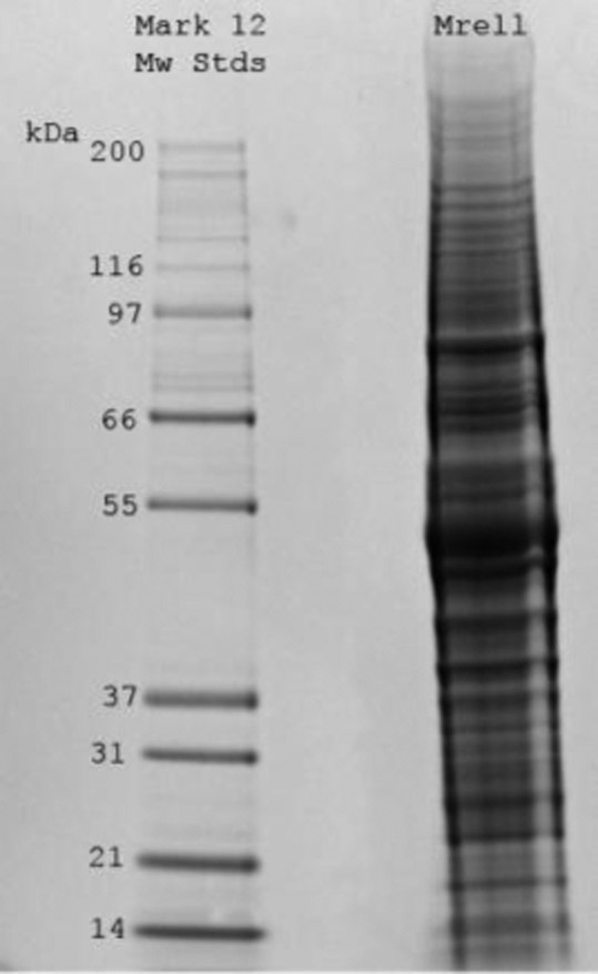
Fig. S1. SDS-PAGE gel of MRE11 immunoprecipitate analysed by mass spectroscopy.
Fig. S2. DNA removal by MRE11 and control immunoprecipitates. (A) Reduction in Hoechst signal by each IP in TARDIS slides probed for topoisomerase IIα. (B) Reduction in Hoechst signal by each IP in TARDIS slides probed for topoisomerase IIβ.
Fig. S3. Removal of topoisomerase II adducts by recombinant MRE11 is dependent on divalent cations. K562 cells were treated with 100 µM etoposide for two hours prior to embedding in agarose on microscope slides. The slides were incubated with MRE buffer, recombinant MRE11 in MRE buffer in the presence or absence of EDTA. Slides were subsequently probed for topoisomerase IIα (A) or IIβ (C) by quantitative immunofluorescence, and Hoechst fluorescence was measured for the same slides (B,D).
Fig. S4. Truncated MRE11 expressed in ATLD cells is distributed differently in the nucleus than WT MRE11. Control and etoposide-treated A-TLD hTERT (expressing only truncated MRE11) and A-TLDwtMRE11 cells were immunostained for MRE11. DNA was visualized with DAPI. For imaging, all MRE11 exposures were kept constant, except for the fifth row, where the exposure time was increased by a factor of 8.