Supplementary material for Fuchs et al. (July 3, 2001) Proc. Natl. Acad. Sci. USA, 10.1073/pnas.121433898
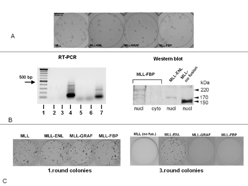
Fig 5.
Control experiments for the CFC test. (A) Titer determination of the various MSCV-MLL-fusion retroviruses on 3T3 cells. Giemsa stained neo-resistant colonies, virus dilution, 10-5. More precisely, the titers were 2.5 × 10⁶ (truncated MLL), 1.6 × 10⁶ (MLL/ENL), 1.8 × 10⁶ (MLL/GRAF), and 1.9 × 10⁶ (MLL/FBP17). (B Left) RT-PCR to ensure mRNA expression of MLL/GRAF (lane 4) and MLL/FBP17 (lane 7) in the myeloid progenitor cells. Because of the possibility that contaminating retroviral cDNA is present in the RNA preparation, the extracts were treated with RNAse-free DNAse for 1 h (Roche Diagnostics) before RT-PCR. After DNAse digestion, the RNA preparations were free of contaminating cDNA. When the RT was omitted, no specific amplification signal for MLL/GRAF (lane 3) and MLL/FBP17 (lane 6) is seen. Only if RT is added to the mixture does a MLL/GRAF and a MLL/FBP17 fusion product become evident. In lanes 2 and 5, an negative (water-) control is shown. (C) Comparison of the first-round cultures and third-round cultures of transduced bone marrow cells. In the first round, numerous colonies were produced even if the cells were transduced with the truncated MLL construct (Left) or the empty MSCV vector (not shown). However, after the third round, only the MLL/ENL transduced cells formed neo-resistant colonies, whereas almost all other colonies disappeard in the cells transduced with MLL/GRAF or MLL/FBP17.