Supplementary material for Thornton et al. (2001) Proc. Natl. Acad. Sci. USA 98 (16), 9413–9418. (10.1073/pnas.151192098)
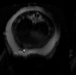
Movie 2.
A series of axial images of a northern elephant seal pup during min 1 of a forced dive. Images are obtained sequentially from the level of the diaphragm through to the pelvic region; splenic margin is outlined in white. Note the change in the splenic tissue, which now appears as a dark crescent due to release of erythrocytes. Volume of spleen = 1,121 ml. Concurrent with splenic contraction is the appearance of the hepatic sinus (light gray amorphous structure appearing in slice 2 and separating into distinct sinuses as the images progress caudally).