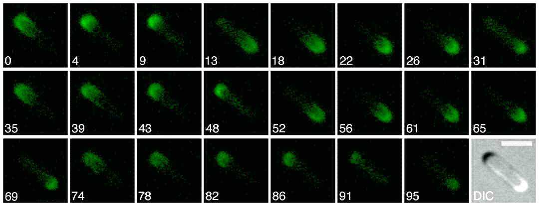
Time-lapse fluorescence micrographs showing the dynamic behaviour of Gfp-MinC. MinCis a division inhibitor which associates, and co-oscillates, with MinD.Pole-to-pole oscillation of MinD, in turn, requires the activity of MinE.On time average, the center of the cell is exposed to the lowest levelof division inhibitor. Times are indicated in seconds. The last panel showsthe cell viewed with DIC (differential interference contrast) optics. The bar in this panel represents 2 m m. Shown is a cell of strainPB114(lDB175)/pDR175[D minCDE(Plac::minDE)/PlR::gfp-minC,cI857].
Cells were grown at 37° C in minimal medium supplemented with 50 mM isopropyl b -d-thiogalactoside, and displayed a normal division pattern. For other details, see Raskin, D. M. & de Boer, P. A. J. (1999) J. Bacteriol. 181, 6419-6424 and Raskin, D. M. & de Boer, P. A. J. (1999) Proc. Natl. Acad. Sci. USA 96, 4971-4976.
2. The MinE pattern

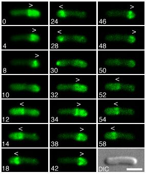
Time-lapse fluorescence micrographs showing the dynamic behavior of MinE-Gfp in normally dividing cells. Times are indicated in seconds. Note the accumulation of MinE-Gfp in the shape of a ring (the E ring) as well as in an extra-annular peripheral pattern (PEA signal), which is present in between the ring and one of the cell poles. Note the net movement of a ring toward the pole with the PEA signal (0-8, 12-28, 32-48, and 52-58 s), the disappearance of a ring and PEA signal when the former approaches a cell pole (8, 28, and 48 s), and the subsequent assembly of a new ring at/near midcell concommitant with the appearance of a new PEA signal in between the new ring and the pole previously devoid of signal (12, 32, and 52 s). Arrowheads (< or >) indicate both the position of the ring as well as the direction of its movement. Shown is a cell of strain HL1/pDR174 [DminDE/Plac::bfp-minD,minE-gfp]. Cells were grown at 30° C in minimal medium supplemented with 37 mM isopropyl b -d-thiogalactoside and displayed a normal division pattern. For additional details, see Hale, C. A., Meinhardt, H. & de Boer, P. A. J. (2001) EMBO J. 20, 1563-1572.