Polar Oscillating MinD Patterns Generated by Traveling Waves of MinE That Remove MinD from the Membrane
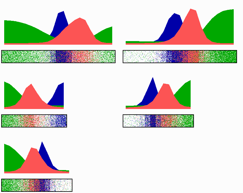
A membrane-associated patterning system (MinE, red) depends on membrane-bound MinD (green) as well as causes its dissociation. In this way, a MinE maximum destabilizes itself, causing its shift toward a region of higher MinD concentration. On its way, the MinE wave removes MinD from the membrane. Shortly before the MinE wave reaches the pole, MinD and MinE concentrations collapse. Meanwhile, a new plateau of membrane-bound MinD is rising in the other part of the cell. A new high MinE concentration is triggered at its flank, causing this peak to disappear also, and so on. A system for septum formation is assumed also to be a pattern-forming system (FtsZ, blue). An inhibition by MinD (green) is sufficient for a reliable localization of the FtsZ pattern in the center of the field. Note that in actual E. coli cells, the inhibition of FtsZ self-assembly is accomplished by the MinC protein that binds to and co-oscillates with MinD.

The following simulation shows first the situation in a large cell. After division, the center is detected rapidly in the smaller cell:
