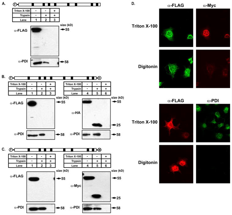
FIGURE 3. The N terminus of DGAT1 has a cytosolic orientation, and the C terminus is exposed to the ER lumen as determined by protease protection analyses.
A, protease protection analysis is shown. Total membranes from HEK-293T cells expressing FL-DGAT1, FL-DGAT-HA, and FL-DGAT-Myc were prepared as described under “Experimental Procedures.” Aliquots of membranes (50 μg protein) were incubated in the absence or presence of 20 μg/ml of trypsin with or without 1% Triton X-100. Samples were immunoblotted with anti-FLAG, which recognizes the N terminus of DGAT1 (A) and anti-Myc (B) or anti-HA (C) that recognize the C terminus of DGAT1. The integrity of membrane vesicles was assessed by immunoblotting with an anti-PDI antibody. D, orientation of the N and C termini of DGAT1 by immunofluorescence microscopy is shown. COS-7 cells transiently expressing FL-DGAT1-Myc were fixed with 4% paraformaldehyde. Fixed cells were incubated with 0.2% Triton X-100 to permeabilize all cellular membranes or 10 μg/ml digitonin to selectively permeabilize the plasma membrane. Cells were exposed to anti-FLAG and anti-Myc antibodies and appropriate secondary antibodies to visualize the N-terminal FLAG (green) and the C-terminal Myc (red) epitopes of DGAT1, respectively (upper panel). Cells were exposed to anti-FLAG and anti-PDI antibodies and appropriate secondary antibodies to visualize the N-terminal FLAG (red) and PDI (green) in the ER lumen. Cells chosen for image comparison had similar levels of expression, as assessed by FLAG signal intensity (lower panel).

