
FIGURE 6. Proposed model of the membrane topology of murine DGAT1.
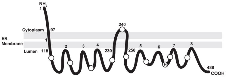
Our topology studies demonstrate that DGAT1 contains three potential transmembrane domains with the N terminus oriented toward the cytosol and the C terminus oriented toward the ER lumen. An alternative model, also consistent with the current data, would show that amino acids 230 –250 do not span the lipid bilayer but are instead embedded within the membrane. H indicates the putative active site histidine at position 426. Circles represent the location of internal Myc or HA epitope tags. Amino acid numbers of regions exposed to the cytosol or ER lumen are indicated.