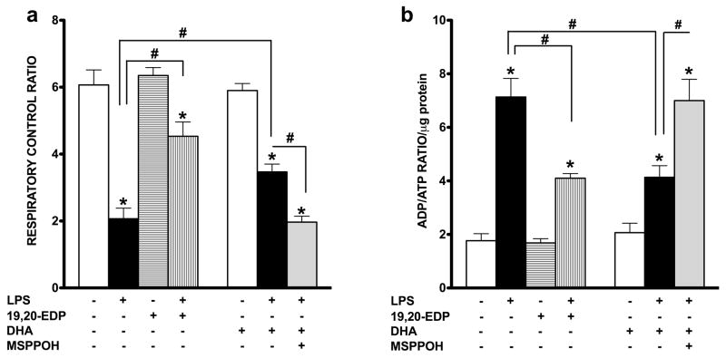
Figure 6. EDPs preserve mitochondrial function following LPS-induced cytotoxicity.
HL-1 cardiac cells were stimulated with LPS (1 μg/ml) in the presence of 19,20-EDP (1 μM), DHA (100 μM) and/or MSPPOH (50 μM) for 24 h. (a) Cells were harvested and transferred into Clark-electrode based chamber connected to Oxygraph at 30 °C. Rates of respiration were measured in saponin-permeabilized cells using 10 mm glutamate and 5 mM malate as substrates. ADP-stimulated respiration was measured after addition of 1 mM ADP. The rates of respiration are expressed as respiratory control ratio (RCR). (b) The intracellular ratio between ADP and ATP was measured by chemolumenescent assay and normalized per μg protein. Values are represented as mean ± S.E.M; N=3 independent experiments; *, p<0.05 treatment vs. vehicle control, #, p<0.05 treatment group vs. LPS or LPS/MSPPOH.

