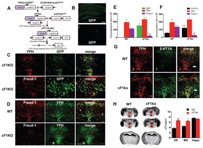
Figure 1. Loss of Freud-1 in 5-HT neurons increases 5-HT1A autoreceptors.
A. Conditional Knockout Strategy. To delete Freud-1 in 5-HT neurons, the cF1ko mouse was generated by crossing CC2D1A(Freud-1)flx/flx mice with TPH2-CreERT2 mice. At 8 weeks of age, mice were administered tamoxifen to activate CreERT2-induced recombination. To delete both Freud-1 and 5-HT1A autoreceptors in 5-HT neurons, the cF1ko mice were mated to the 5-HT1Aflx/flx mice, in which the 5-HT1A gene is flanked by LoxP sites and a YFP cassette to generate the Freud-1/5-HT1A double knockout mice following tamoxifen administration. B. Tamoxifen-induced recombination specificity. Hippocampal and prefrontal cortex sections from tamoxifen-treated conditional Freud-1 knockout/ROSA-GFP mice (cF1KO) show background GFP staining. C. Tamoxifen-induced recombination and loss of Freud-1 in dorsal raphe. DR sections from tamoxifen-treated cF1KO mice were stained for GFP and either TPH or Freud-1 (10× magnification, scale bar = 20 μm; inset, 20x). GFP was present in 92% of TPH+ cells, while 88% of Freud-1 was in GFP-cells in cF1KO sections (n=4). D. Loss of Freud-1 in 5-HT neurons. Freud-1/TPH-labeled cells in DR were almost absent in cF1ko compared to WT. By contrast, Freud-1+/TPH-cells remained (white arrowheads) (n=4). E. Quantification of GFP-, TPH-and Freud-1-stained cells in dorsal raphe of cF1KO and WT mice (n=4), shown as mean ± S.E. (p <0.001). F. Quantification of 5-HT1A-, TPH- and Freud-1-stained cells in dorsal raphe of cF1ko (non-ROSA-GFP) and WT mice (n=4), shown as mean ± S.E. (p <0.001). G. Loss of Freud-1 and increased 5-HT1A-positive cells in dorsal raphe of cF1ko mice. DR sections from tamoxifen-treated cF1ko vs. F1wt (WT) mice (scale=50 μm, n=4) were stained for TPH and 5-HT1A receptors (arrow, 5-HT1A in TPH-cells); 5-HT1A receptors were increased in TPH+ cells. H. Increased 5-HT1A binding in raphe of cF1ko mice. At left are representative images of 125I-MPPI autoradiography of sections from cF1ko and WT mice in dorsal and median raphe (boxes) at two levels (Bregma −4.60 and −4.72 mm) and hippocampus (Bregma −1.70). At right is quantification of 125I-MPPI binding. Data represent mean ± SEM (n=4/group), *p < 0.05; **p < 0.01. 5-HT1A binding was increased in raphe of cF1ko mice; DR (unpaired two-tailed Student’s t-test, DF=6, t=9.129, **p<0.001), MR (unpaired two-tailed Student’s t test, DF=6, t=6.635 *p<0.01).

