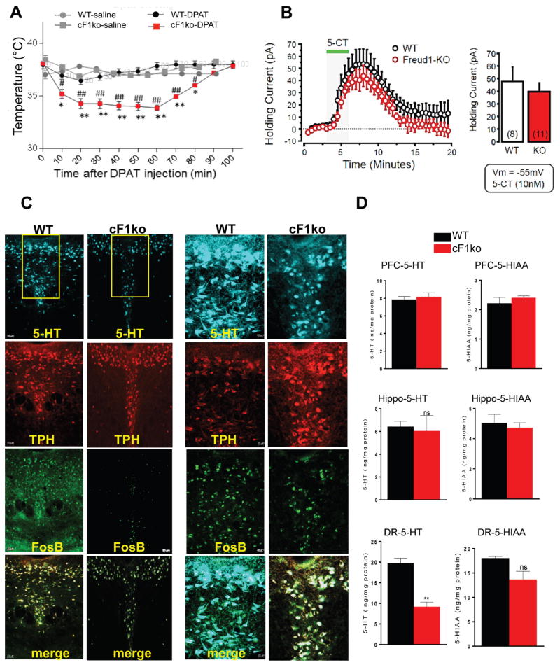
Figure 2. Loss of Freud-1 augments 5-HT1A autoreceptor function and reduces 5-HT neuron activity and 5-HT levels.
A. 5-HT1A-induced hypothermia. 8-OH-DPAT (0.75 mg/kg, i.p.) induced a greater body temperature reduction in cF1ko compared to F1wt (WT). Data represent mean±SEM (n=3/group); *p<0.05 vs. WT-saline, **p<0.001 vs. wild-type-DPAT, #p<0.05 vs. cF1ko-saline, ##p<0.001 vs. cF1ko-DPAT. B. Whole-cell voltage-clamp recordings (Vm = −55 mV) of 5-HT neurons in slices of dorsal raphe in vitro, from cF1ko or F1wt mice (n = 4) in response to 5-CT (10 nM). No significant difference in 5-HT1A receptor-induced outward current was observed. C. Reduced 5-HT- and FosB-stained cells in cF1ko raphe. DR sections of cF1ko or F1wt (WT) mice stained for 5-HT, TPH, and FosB shown at 10× (left, scale=50 μm) or 20× magnification of boxed region (right, scale=20 μm). D. Reduced raphe 5-HT content in cF1ko mice. Tissue 5-HT and 5-HIAA content was quantified by HPLC for dorsal raphe, hippocampus (Hippo) and prefrontal cortex (PFC) of cF1ko vs. WT mice. Data represent mean ± SEM, n=3/group; reduced raphe 5-HT content in cF1ko vs. WT mice (unpaired two-tailed Student’s t test, DF=4, t=6.675 **p<0.01).

