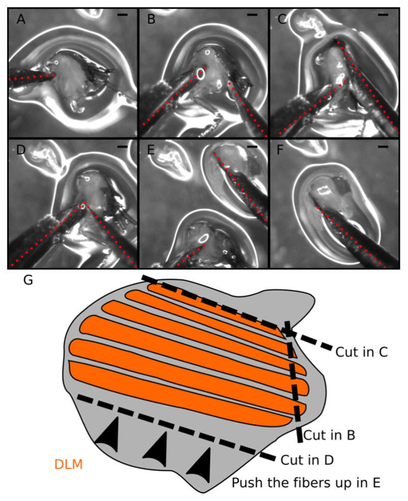
Figure 4. Step-by-step guide to dissect DLM fibers.

A–F. Snapshots from Video 2. A. Locate and hold the half-thorax using the left needle. Place the needle just below the DLM and above the legs. B. Make a small incision at the DLM posterior attachment site. C. Make a long cut above the DLM to completely separate the muscle fibers from the dorsal cuticle. D. Move the right needle to the ventral DLM (near the left needle). E. With the right needle push the DLM fibers out of the thorax. F. Isolated DLM can be further separated into individual fibers. G. Cartoon illustrating the dissection process. Dotted red lines mark the dissection needles. Note the small amount of liquid (~100 μl) in the dissection plate to prevent floating of half-thoraces. Scale bars = 200 μm.
