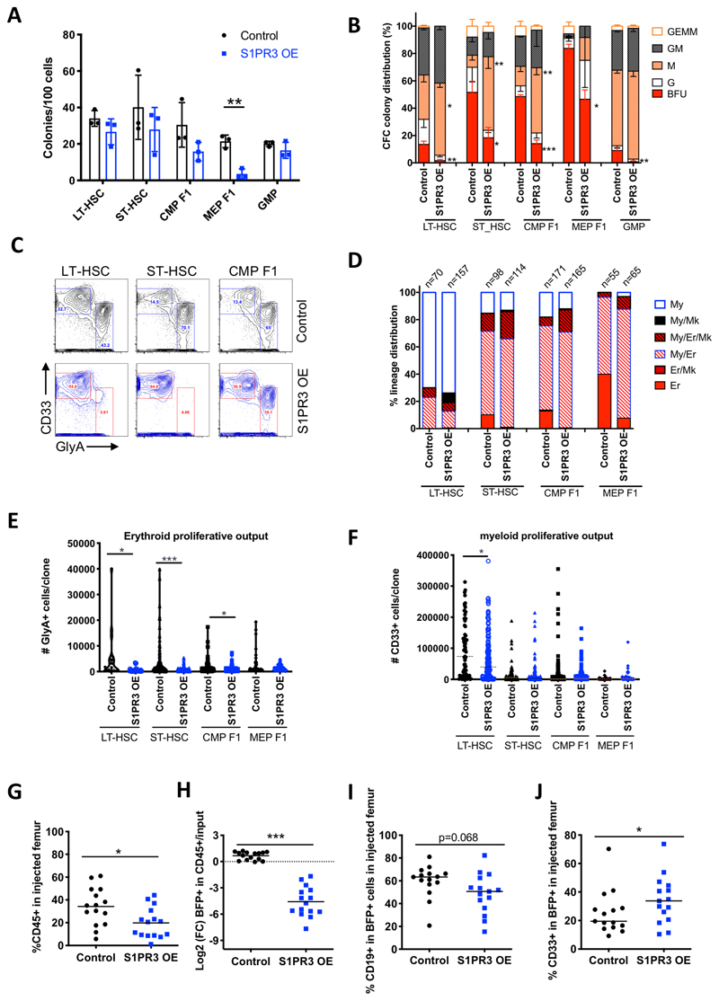
Figure 2. S1PR3 overexpression is sufficient to promote myeloid differentiation in human HSPC in vitro and in vivo.
A, The number of colonies for 100 transduced cells from the indicated populations at 10 days of a CFC assay for control and S1PR3OE lentiviral knockdown vectors (n=3). B, Normalized colony distribution showing burst-forming units (BFU) are significantly decreased with S1PR3OE and macrophage (M) colonies are significantly increased from all scored cell populations (n=3 CB). G (granulocyte); GEMM (granulocyte erythrocyte monocyte megakaryocyte). C, Myeloid (CD33+) vs erythroid (GlyA+) lineage distribution for all pooled cells from one representation assay at day 14 from (A). D, Lineage distribution outcomes in single cell assays performed on MS5 stroma in erythroid-myeloid cytokine conditions. Data for indicated number of cells scored in each condition pooled from 3 CBs. E-F, Average number of GlyA+ or CD33+ cells from single cell differentiation assays (D). G, Human CD45 chimerism, H, Log2 fold change (FC) in BFP which marks transduced cells relative to input, I, CD19+ B lymphoid cells in the transduced fraction and J, CD33+ myeloid cells in BFP+ cells for control or S1PR3OE measured in the injected femur at 4 weeks post-transplant in xenotransplantation, 5 mice for each condition from 3 CB. *** P<0.001** p<0.01, *<0.05, unpaired Student t-test. Data are mean and s.d. except for E-F where data are mean and s.e.m.

