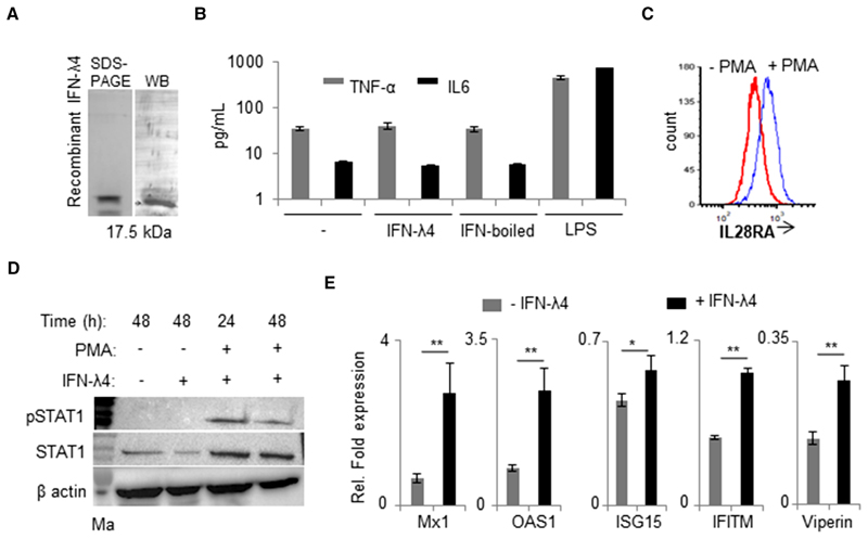
Figure 1.
IFN-λ4 can signal in monocytes after PMA treatment. (A) Western blot analysis of the human recombinant IFN-λ4 used in the study. (B) TNF-α and IL-6 cytokines secretion was measured by ELISA from cell-free supernatants of THP-1-derived macrophage-like cells treated for 48 h with1 μg/ml IFN-λ4, boiled IFN-λ4 preparation, or 1 μg/ml of LPS; untreated macrophages served as control; mean and error bars depicting SD from technical replicates are shown. (C) THP-1 cells were differentiated into macrophage-like cells by treatment with PMA (100 nM) for 48 h. The cells were removed and compared with fresh untreated THP-1 cells for surface expression of IL28RA using flow cytometry. (D) pSTAT1 and STAT1 expression was assessed by Western blot in THP-1 cells treated or not with PMA (100 nM) in the presence or absence of IFN-λ4 (1 μg/ml) for 24 and 48 h as indicated. α-actin was used as loading control. The presented immunoblot is a representative image of two independent experiments. derived macrophage-like cells treated or not with 5 μg/ml of IFN-λ4 for 24 h. The data are representative of two independent experiments showing (E) IFN-stimulated genes expression was analyzed by quantitative polymerase chain reaction (qPCR) based on RNA samples extracted from THP-1mean of technical replicates from a single experiment with SD depicted by error bars. *P < 0.05; **P < 0.01

