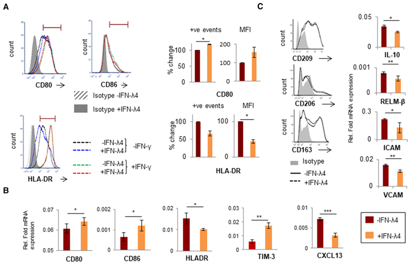
Figure 3.
IFN-λ4 confers a mixed phenotype on THP-1-derived macrophage-like cells. (A) Histogram from one representative experiment showing that IFN-λ4-treated macrophage-like cells had increased expression of CD80 but reduced expression of HLA-DR (left) when activated with LPS, whereas IFN-λ4 had no effect when activation was carried out with LPS and IFN-λ. The graph on the right shows the normalized (for mock IFN-λ4 treatment; error bars show SD. The significance was calculated using a one-sample t-test (*P < 0.05). (B and C, right) mRNA expression of diftreatment) mean of positive events in the gate shown in the histogram and/or mean fluorescence intensity from two independent experiments with ferent genes from M1-activated (with LPS) (B) and M2-activated (with macrophage CSF [M-CSF], IL-4, and IL-10) (C) THP-1-derived macrophagereplicate experiments. *P < 0.05; **P < 0.01, ***P < 0.001. (C, left) Histogram of CD209-, CD206-, and CD163-positive cells as determined by flow like cells. The data show the mean from technical triplicates with error bars depicting SD, and are representative of at least two separate biological cytometry of THP1-derived M2 macrophage-like cells differentiated in the absence or presence of IFN-λ4 (6 μg/ml). The data were obtained from a single experiment

