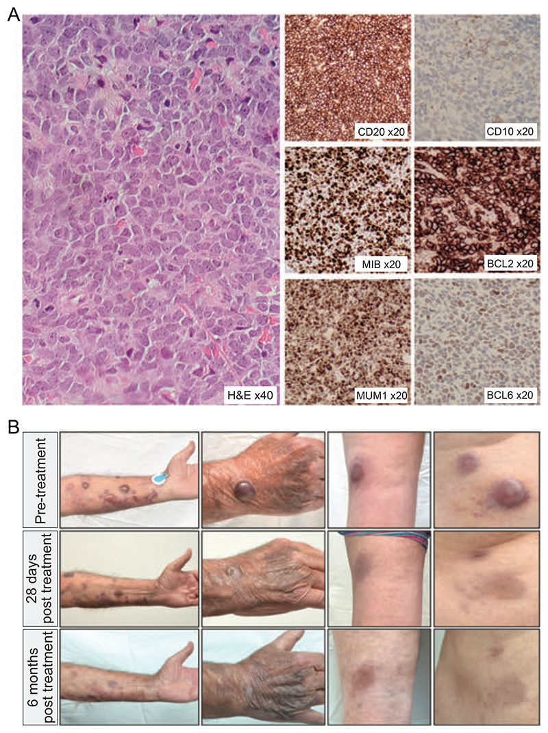
Figure 1.
(A) Histology and immunohistochemistry from tumor biopsy showing a diffuse infiltration by large atypical cells with predominantly centroblast-like morphology expressing CD20, MUM1, BCL2, weak BCL6, no CD10 and MIB1 proliferation faction 90% (B) Images of selected lesions on the forearm, hand, popliteal fossa and abdomen taken prior to therapy (top row), at 28 days (middle row) and at 6 months (bottom row) of BCR-targeted therapy.

