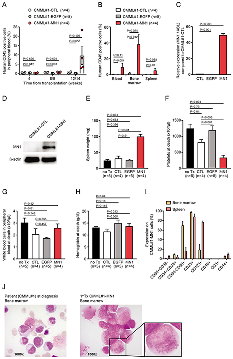
Figure 1. Supplementation of primary CMML cells with MN1 promotes engraftment in vivo.
A) Engraftment of human hematopoietic cells (CD45 positive) in the peripheral blood of primary recipient NSGS mice monitored at the indicated time points. Recipient mice were transplanted with untransduced CMML#1 cells (CMML#1-CTL), EGFP- (control vector) transduced CMML#1 cells (CMML#1-EGFP) or MN1-transduced CMML#1 cells (CMML#1-MN1), (number of analyzed mice is indicated in the figure; mean ± SEM).
B) Engraftment of human CD45 positive cells in peripheral blood, bone marrow and spleen of primary recipient NSGS mice at sacrifice (number of analyzed mice is indicated in the figure; mean ± SEM).
C) Expression levels of MN1 in untransduced CMML#1 patient cells (CMML#1-CTL), EGFP-transduced CMML cells (CMML#1-EGFP) and MN1-transduced CMML cells (CMML#1-MN1). Gene expression was determined by quantitative RT-PCR relative to the housekeeping gene ABL1 and normalized to gene expression in untransduced CMML#1 patient cells (n=3), (mean ± SEM).
D) Western blot showing MN1 expression in primary bone marrow mononuclear cells from a patient with CMML (CMML#1-CTL) and in their MN1-transduced counterparts isolated from bone marrow of engrafted recipient mice. ß-actin was used in the same blot as loading control.
E) Spleen weight of primary recipient NSGS mice at sacrifice (number of analyzed mice is indicated in the figure; mean ± SEM).
F) Platelet count in peripheral blood of primary recipient NSGS mice at sacrifice (number of analyzed mice is indicated in the figure; mean ± SEM).
G) White blood cell count in peripheral blood of primary recipient NSGS mice at sacrifice (number of analyzed mice is indicated in the figure; mean ± SEM).
H) Hemoglobin level in peripheral blood of primary recipient NSGS mice at sacrifice (number of analyzed mice is indicated in the figure; mean ± SEM).
I) Immunophenotype of engrafted CMML#1 cells from bone marrow and spleen of primary recipient NSGS mice at sacrifice (gated on human CD45+EGFP+ CMML#1-MN1 cells; n=3; mean ± SEM).
J) Morphology from bone marrow smears of the CMML#1 patient at diagnosis and from a transplanted mouse at sacrifice. The enlarged cell shows granules in the cytoplasm that can be identified as lipid granules in cells from later transplantations (see Figure 3B).

