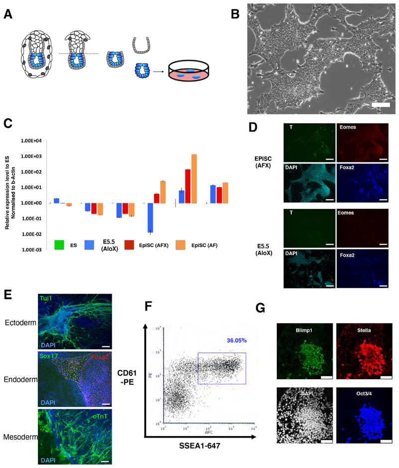
Figure 1. Derivation of stem cell lines from formative epiblast.
(A) Schematic of cell line derivation from E5.5 epiblast. (B) Image of serially passaged E5.5 epiblast-derived culture. Scale bar 100μm. (C) RT-qPCR analysis of marker gene expression in AloX cells and EpiSCs relative to ES cells in 2iL (=1), normalized to beta-actin. Error bars are S.D. from technical triplicates. (D) Immunofluorescent staining of EpiSCs and AloX cultures for early lineage markers. Scale bars 150μm. (E) Immunostaining of embryoid body outgrowths for germ layer markers, DAPI in blue. Scale bars, 150μm. (F) Flow cytometry analysis of PGCLC induction at day 4. (G) Immunostaining of day 4 PGCLC. Scale bars 50μm.

