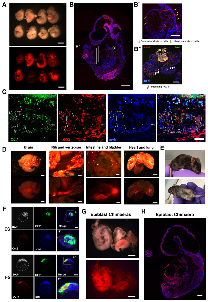
Figure 3. Blastocyst chimaera contribution by FS cells and formative epiblast.
(A) Bright field and fluorescent images of E9.5 embryos generated after blastocyst injection of mKO2 reporter FS cells. Scale bar is 1 mm. (B) Sagittal section from one chimaera, stained for mKO2 and DAPI. Inset B’, mKO2 positive cells in foregut endoderm (yellow arrowheads) and cardiac mesoderm (green arrowheads). Inset B” (rotated 900), Sox2 immunostaining (white arrowheads) in the hindgut region. Scale bars, 200μm (B), 100μm (B’, B”). (C) mKO2 positive cells expressing Oct4 and Mvh PGC markers in E12.5 chimaeric gonad. Triple positive cells are highlighted with dashed circles. Scare bars, 75μm. (D) Fluorescent images of organs from post-natal (P21) chimaera overlaid with 20% opacity bright field image. Scale bars, 2 mm. (E) Coat colour chimaera at P14. (F) Blastocysts injected with GFP reporter ES cells or FS cells and cultured for 24 hours. ES cells are Klf4+Oct6- (n=11) (F’) whereas FS cells are Klf4-Oct6+ (F”) (n=15). Scale bars, 40μm. (G) E9.5 chimaeras obtained from blastocyst injection of mTmG expressing E5.5 epiblast cells. Scale bars, 500μm. (H) Section from left embryo in Panel G stained with anti-RFP to visualise membrane-tdTomato, DAPI in blue. Scale bar, 200μm.

