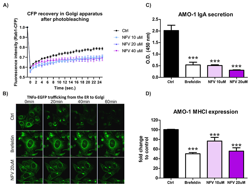
Figure 3. Nelfinavir impairs intracellular trafficking, plasma membrane deposition and secretion of ER-resident proteins.
A) FRAP of CFP-Rab1A protein in the Golgi of control, untreated cells or cells pretreated for 3h with increasing doses of nelfinavir. B) Representative picture of TNFα-eGFP retained in the ER of the U-2 OS cells after 3h treatment with 10 μM brefeldin A or 20 μM nelfinavir. For the movies showing trafficking of TNFα-eGFP after the treatment see Movie S1A-C. For the quantification of TNFα-EGFP signal retained in the cell after exposure to nelfinavir and other drugs see Figure S6. C) IgA secretion in AMO-1 MM after the treatment for 3h with 10 μM brefeldin A or 10 μM and 20 μM nelfinavir. D) Surface expression of MHC class I on AMO-1 cells after the treatment for 3h with brefeldin A or 10 μM and 20 μM nelfinavir. Data for C and D represent means ±SD from three independent replicates, statistically significant differences are marked with *** at p<0.001.

