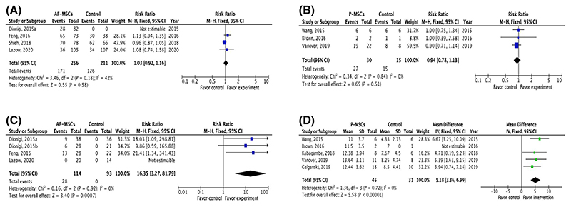
Figure 3. Meta-analysis.
(A) Meta-analysis of fetal rat survival at term after intra-amniotic injection of allogenic amniotic fluid-derived mesenchymal stem cells or saline at E17.37,39–41 Myelomeningocele (MMC) was created in all studies using retinoic acid. (B) Meta-analysis of fetal lamb survival at term after application of human second trimester placental (P)-mesenchymal stem cells (MSCs) during fetal surgical closure of MMC compared to fetal surgical closure alone.48,49,52 MMC was surgically created in these studies at Gestational Age (GA) 75–77 days; fetal surgical closure was performed 25 days later (GA 100–102 days). (C) Meta-analysis of defect coverage in the retinoic acid-induced fetal rat MMC model. Intra-amniotic injection of allogenic amniotic fluid-derived mesenchymal stem cells at E17 significantly increased the likelihood of total defect coverage compared to saline injection.37–39,41 (D) Meta-analysis of spinal cord function in the surgical fetal ovine model of MMC determined by sheep locomotor rating scale, after fetal surgery in conjunction with the application of human placental-derived mesenchymal stem cells compared to fetal surgery alone48–50,52,53 [Colour figure can be viewed at wileyonlinelibrary.com]

