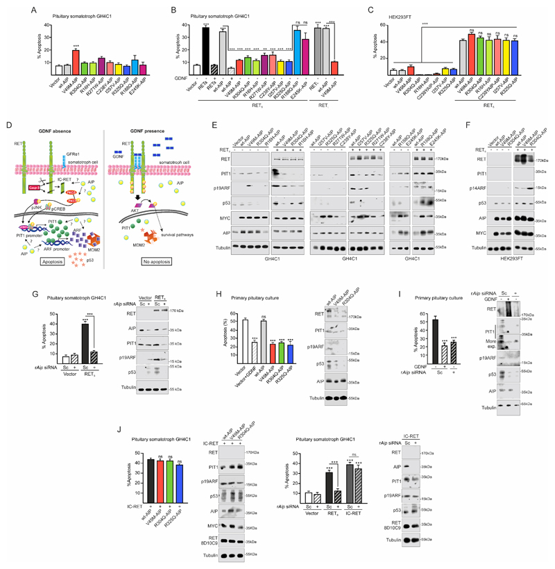
Figure 2. Variant AIP or Aip downregulation blocks the physiological RET/PIT1/CDKN2A-ARF/p53 apoptosis pathway.
A) Expression of wt or variant AIP does not affect basal rate of apoptosis when GH4C1 cells are deprived of GDNF (0.5% FBS). V49M is the only variant causing some apoptosis. B) When RETS is transfected in GH4C1, serum deprivation induces the RET-apoptotic pathway (black bar) that is blocked by the RET ligand GDNF (black). Expression of human wtAIP together with RET maintain the apoptotic pathway which is also blocked by GDNF (grey colour, black). Expression of variant AIP (R16H, V49M, R271W, C238Y, I257V, R325Q, coded coloured bars) blocks RET-induced apoptosis in the absence of GDNF. RETL expression follows the same pattern with strong apoptosis in the presence of wtAIP (grey bar) blocked by variant AIP in the absence of GDNF. Expression of R188Q or E245K (coded coloured bars), does not block RET-induced apoptosis. C) A similar experiment in the embryonic kidney cell line HEK293T cells have different results. RET induces apoptosis in the presence of wtAIP but also in the presence of any AIP variant. D) Cartoon showing the known RET pathways in pituitary somatotroph cells: left, RET/PIT1/CDKN2A-ARF/p53 apoptosis pathway; right, RET/GDNF-AKT survival pathway. AIP alteration could affect any step in the apoptotic pathway or directly induce the survival pathway. E) AIP variants that block apoptosis also prevent RET-induced Pit1 overexpression, p19Arf increase and p53 accumulation. The two AIP variants that did not block apoptosis did not alter RET-induced Pit1 overexpression, p19Arf increase and p53 accumulation. F) HEK293T does not express PIT1 and express large T-antigen. However, RET expression induces ARF and p53 accumulation in the presence of WT or variant AIP. G) In GH4C1 repression of endogenous Aip blocks RET-induced apoptosis in parallel to PIT1, ARF and p53 repression. H-I) Similar results were obtained in rat male primary pituitary culture with 45%-50% somatotroph cells expressing endogenous rat RET receptor. H) When cultures are serum deprived (no GDNF) apoptosis is induced, and this is prevented by addition of rGDNF. Transfection of wtAIP does not alter apoptosis, nor the apoptotic pathway through PIT1/ARF/p53 accumulation. Transfection of variant AIP blocks PIT1/ARF/P53 increase and apoptosis. I) Repressing Aip with siRNA blocks PIT1/ARF/p53 increase and apoptosis as well as GDNF addition. J) In GH4C1, direct transfection of the short caspase-processed intracellular RET fragment, IC-RET, induces the RET/PIT1/CDKN2A-ARF/p53 apoptosis pathway that cannot be blocked by AIP mutants or rAip siRNA.
(One-way ANOVA with Tukey’s multiple comparison test correction A, J left; Two-ways ANOVA with Sidak’s multiple comparison test correction B-C-G-H-I-J right. **, p<0.01; ***, p<0.001; ns, non-significant).

