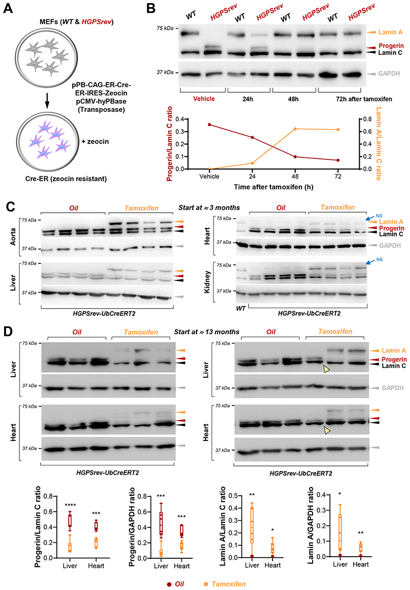
Figure 6. In vitro and in vivo tamoxifen-induced Cre-dependent progerin suppression and lamin A restoration.
(A) Wild-type (WT) and HGPSrev mouse embryonic fibroblasts (MEFs) were co-transfected with plasmids to confer resistance to zeocin and express a tamoxifen-inducible Cre recombinase. (B) Zeocin-resistant MEFs were analyzed by western blot to examine lamin A/C, progerin and GAPDH expression. Equal volumes of ethanol or tamoxifen (25 nM final concentration) were added to the cells as indicated. The graph shows the relative amount of progerin and lamin A in HGPSrev MEFs (normalized to lamin C content). (C, D) Western blot analysis of tissues of LmnaHGPSrev/HGPSrev Ubc-CreERT2-tg/+ mice which received vehicle (oil) or tamoxifen beginning at the age of ≈3 months (C, n=4 each group) and ≈13 months (D, n=6 each group). Mice in C were euthanized 1 week after oil or tamoxifen administration, and mice in D when they met human end-point criteria. Yellow arrowheads in D indicate one animal in which tamoxifen administration did not suppress progerin or induce lamin A and that died 2 days after the end of tamoxifen administration (see Figure 7B, bottom right). Quantification of the relative amounts of lamin A and progerin in the blots in C is shown in Supplemental Figure S5). The graphs in D show the relative amount of progerin and lamin A normalized to lamin C and GAPDH content. Statistical analysis to compare genotypes was performed by two-tailed t-test. *, p<0.05; **, p<0.01; ***, p<0.001; ****, p<0.0001. Each symbol represents 1 animal. NS in panel C indicates nonspecific band.

