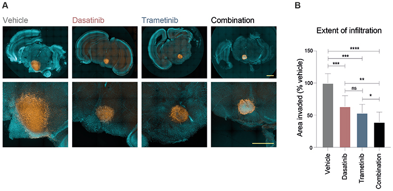
Figure 7. Efficacy of combined dasatinib and trametinib on ex vivo brain slice preparations.
(A) Coronal slices of normal mouse brain, counterstained with Hoechst33342 (aqua) are implanted in the pontine region with ICR-B169 parental cells, stained with human nuclear antigen (orange), and treated for 4 days with 1μM dasatinib, 0.1234μM trametinib, or both, compared to vehicle control. Scale bar is 2 mm. (B) Barplot of quantification of tumour cell infiltration across the brain parenchymal tissue as measured by the calculated area invaded compared to vehicle control. Plotted is the mean of at least 6 independent slices, error bars represent the standard deviation. ****p<0.0001, ***p<0.001, **p<0.01, *p<0.05, FDR-corrected t-test.

