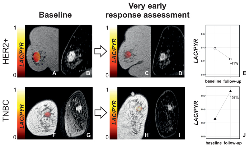
Figure 1. Changes in LAC/PYR between baseline and very early response assessment in a responder and non-responder.
(A,C,F, and H) Coronal T1-weighted 3D spoiled gradient echo (SPGR) images with LAC/PYR map overlaid on the breast tumor. (B,D,G, and I). Coronal reformatted DCE images obtained 150 s after i.v. injection of a gadolinium-based contrast agent. A patient with HER2+ breast cancer was imaged at baseline (A and B) and for ultra-early response assessment (C and D) following standard-of-care treatment and showed a decrease in LAC/PYR of 41% (E) indicating non-response. At surgery, non-pathological complete response (non-pCR) with residual invasive cancer was identified. Another patient with TNBC was imaged at baseline (F and G) and for ultra-early response assessment (H and I) following treatment with chemotherapy and a PARP inhibitor and showed an increase in LAC/PYR of 157% (J) indicating response. At surgery, pathological complete response (pCR) without residual invasive breast cancer was found. TNBC: triple-negative breast cancer; HER2+: HER2/neu (human epidermal growth factor receptor 2) positive.

