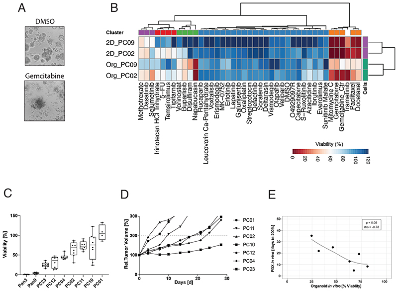
Figure 2. Drug response in PDAC organoids and PDX models are correlated.
(A) Brightfield images for the PC02 PDAC organoid line, treated with DMSO control (upper panel) and the standard PDAC drug gemcitabine (1μM) (lower panel). (B) Drug response profile of paired isogenic PDAC organoid lines and corresponding monolayer cell lines (PC02 and PC09). Tested drugs are either approved for PDAC by the FDA or currently in clinical trials. Viability was normalized to solvent control (0.1% DMSO) and at a reference dose of 1μM (red = high sensitivity, blue = low sensitivity). Data displayed are averages of the technical and biological replicates. (C) Relative viability (compared to DMSO control) of WT pancreas organoid lines and PDAC organoid lines treated with 10 μM erlotinib. Technical replicates of two independent experiments are shown as Tukey plots. (D) Mean relative tumor volume of the same PDAC lines as shown in (c) grown as PDX models and treated with erlotinib (d=day, n=5 mice per group). (E) Correlation of drug responses for erlotinib in vitro (% viability at a dose of 10μM) and in vivo (Days to reach 200% tumor volume) using a Spearman’s rank correlation test. See also Figure S2.

