Figure 3.
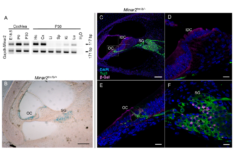
Expression of Minar2, localization of the protein in the cochlea and innervation of cochlear hair cells in Minar2 heterozygous mice, using the LacZ reporter gene component of the inserted cassette in the mutant allele which expresses β galactosidase (see Appendix SI, Fig. S3). (A) RT-PCR of Minar2 expression in the cochlea at embryonic 18.5 (E18.5), postnatal day 0 (P0), and day 30 (P30). Also, expression in different mouse tissues at P30 like Hc: hippocampus, Cx: cortex, Li: liver, Sp: spleen, Ki: kidney, Lu: lung. Gapdh was used as a control. (B) Localization of Minar2 using the reporter gene LacZ of the mutant allele and in β-gal staining shown in blue. Note the localization of Minar2 at the SG: spiral ganglion, OC: organ of Corti. Scale bar: 70 µm. (C) Cross section from 24 kHz region of P1 mutant inner ear was labelled with anti-β gal (magenta) to detect Minar2 localization; and Tuj1 (green) to label neurons. Bar=70µm in C and a zoom in is shown in (D), (E), and (F) scale bar=10µm. SG; Spiral ganglion, OC: organ of Corti, IDC: Interdental cells.

