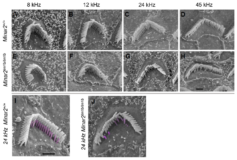
Figure 4.
Scanning electron microscopy reveals stereocilia defects. Outer hair cells at 8kHz (85% of distance from base), 12kHz (70%), 24kHz (40%) and 45kHz (20%) best frequency locations in wildtype mice (A-D) and Minar2tm1b homozygotes (E-H). Higher magnification images with the shortest row colored in magenta in a wildtype (I) and a mutant (J) outer hair cell, showing the reduction in numbers. Scale bars (in H and I) indicate 1 μm.

