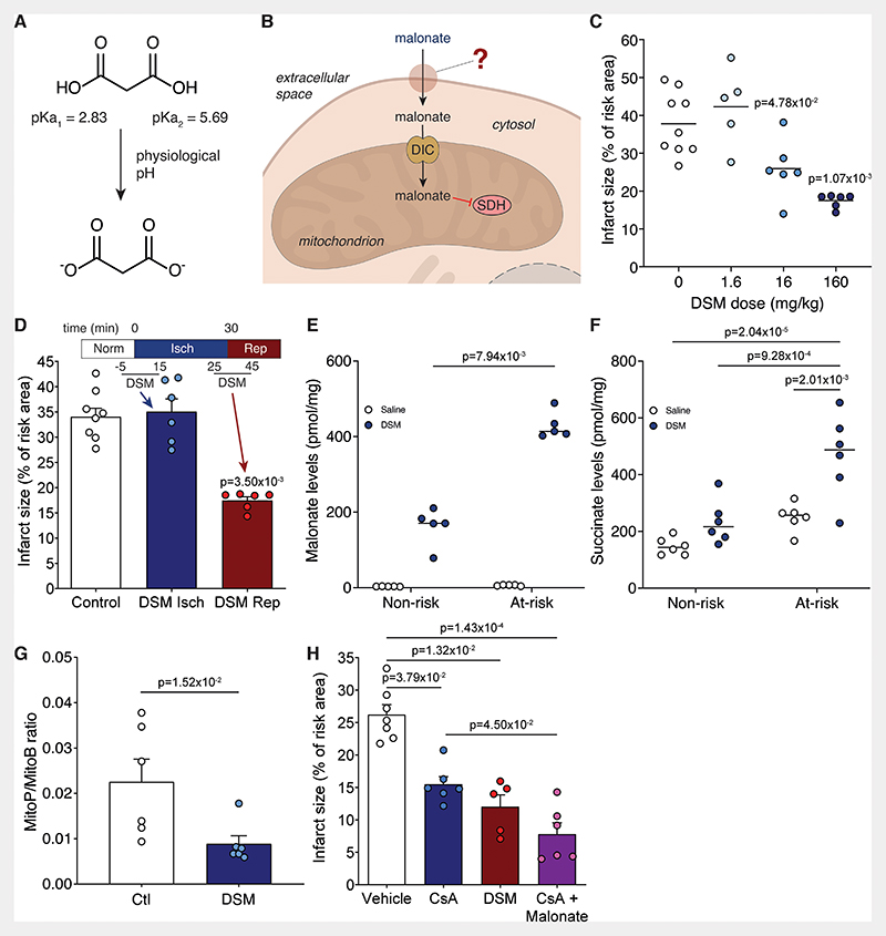
Figure 1. Malonate is cardioprotective only when given at reperfusion.
A, Malonate predominantly carries two negatively charged dicarboxylates at physiological pH. B, Schematic of the barriers to effective malonate delivery. C, Infarct size in murine LAD model with infusion of varying doses of DSM (0, 1.6, 16 or 160 mg/kg; n=9, 5, 6, 6 respectively) infused at reperfusion after 30 min ischemia, quantified by TTC staining. D, Infarct size in murine LAD model with infusion of DSM (160 mg/kg) at ischemia or at reperfusion (mean ± S.E.M., n=8 (Control), 6 (DSM isch and DSM rep) biological replicates. E and F, Levels of malonate (E) and succinate (F) in non-risk and at-risk tissue after 1 min reperfusion with either saline or DSM (160 mg/kg) after 30 min ischemia (mean ± S.E.M., n=5 (E), 6 (F) biological replicates). Statistics: Kruskal-Wallis with Dunn’s post hoc test (C to E), two-way ANOVA with Tukey’s post hoc test (F). G, MitoP/B ratio in at-risk heart tissue from LAD model after 30 min ischemia and 15 min reperfusion with either saline (Ctl) or DSM (160 mg/kg) infusion (mean ± S.E.M, n=6 biological replicates, statistics: unpaired, two-tailed Mann-Whitney U test) H, Infarct size in murine LAD model with infusion of vehicle (ethanol/Cremphor EL in saline) ± cyclosporin A (10 mg/kg) or DSM (160 mg/kg) at reperfusion (mean ± S.E.M, n=7 (vehicle), 6 (CsA, CsA + Malonate), 5 (DSM) biological replicates, statistics: Kruskal-Wallis with Dunn’s post hoc test).

