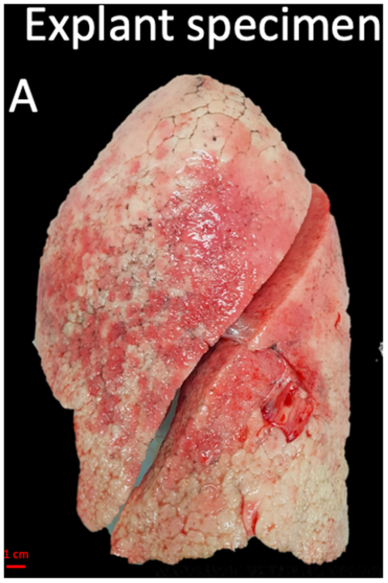
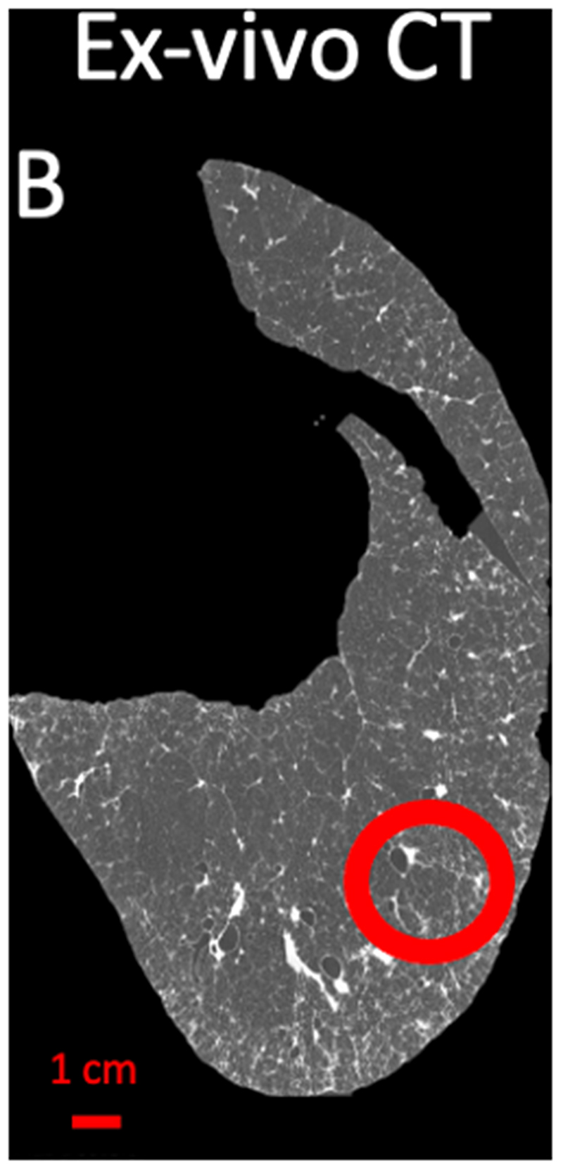
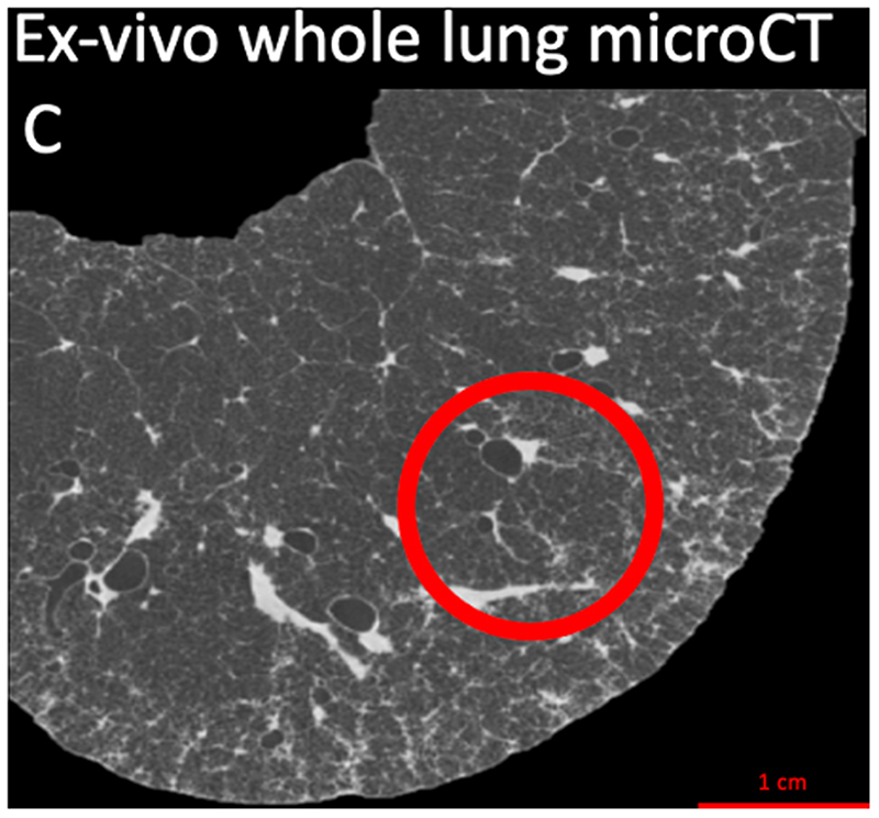
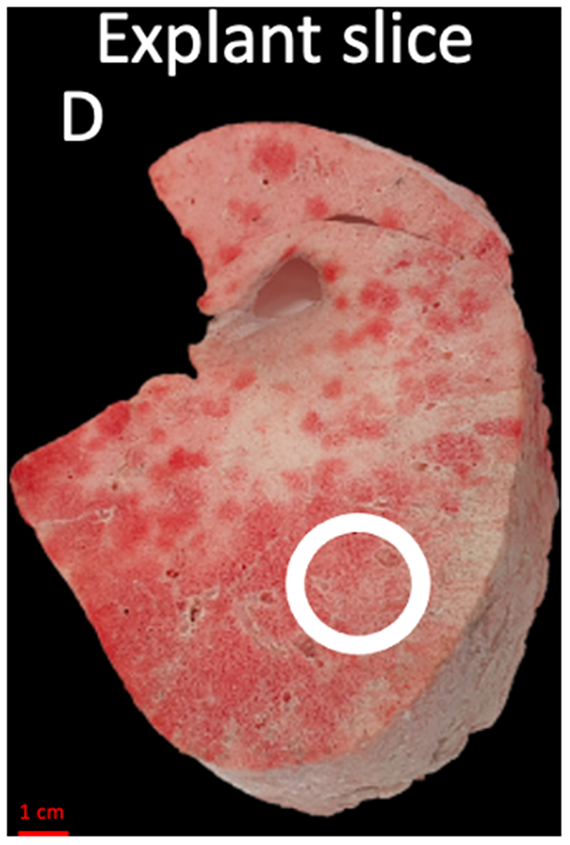
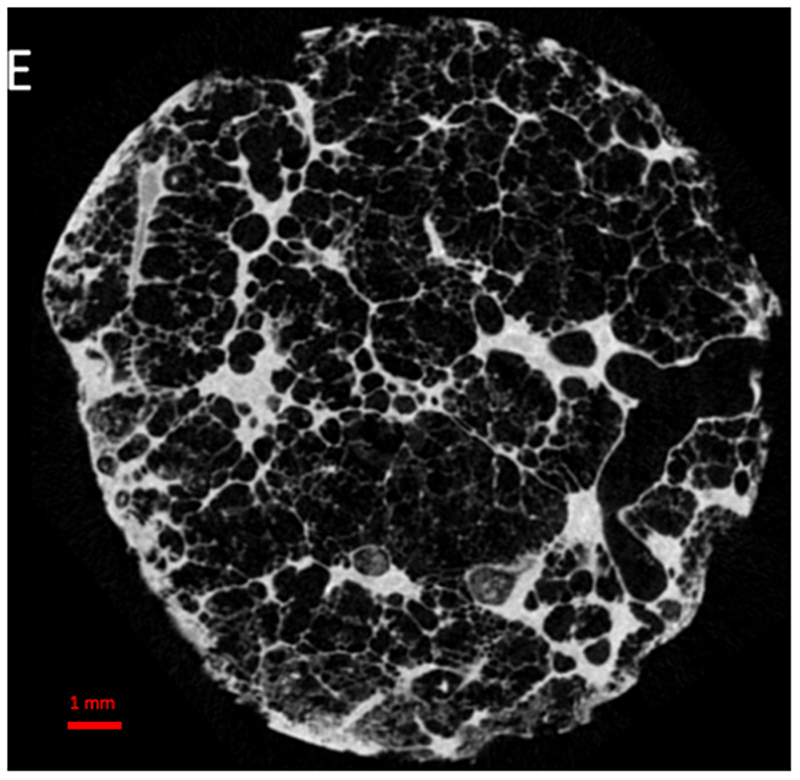
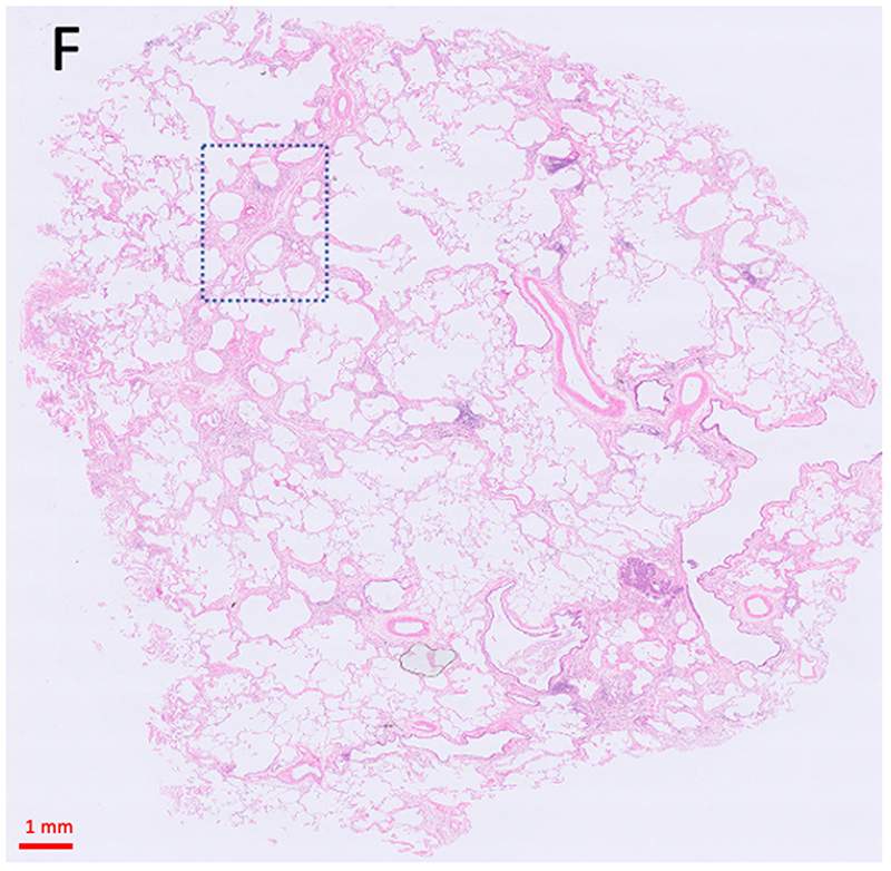
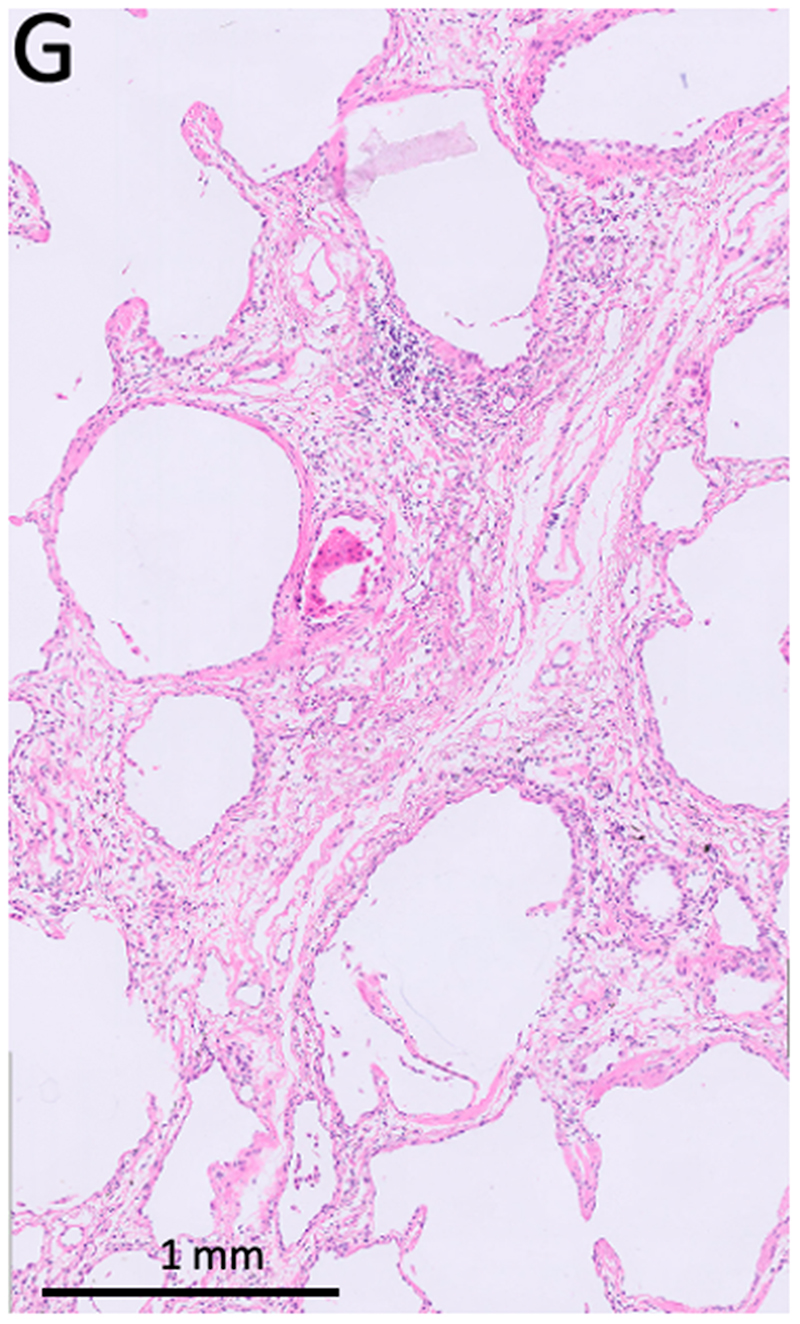
Figure 2. Study design.
(A) The explant lung was frozen solid in liquid nitrogen fumes. (B) An ex-vivo noncontrast CT scan was obtained of the specimen while it was frozen (axial view). (C) For better spatial resolution, a whole lung micro-CT was obtained. (D) The lung was sliced transversally in 2 cm slices. (E) Micro-CT scan of a core sample indicated with the circle in B, C, and D was performed. (F) Matched H&E image (5x magnification) at the same location of the micro-CT scan. (G) High magnification H&E image (20x magnification) view showing para-septal fibrosis

