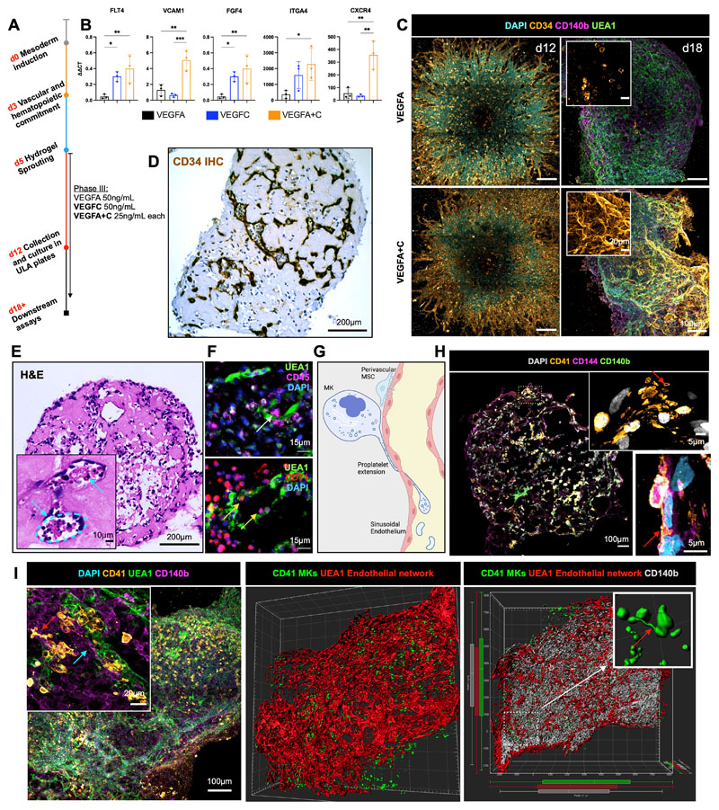
Figure 2. Addition of VEGFC induces specialization of organoid vasculature to a bone marrow sinusoid-like phenotype.
(A) In the sprouting phase of differentiation (D5) in hydrogels, organoids were supplemented with either VEGFA or VEGFC, or both VEGFA and VEGFC. (B) mRNA expression of canonical cell surface receptors, growth factors and adhesion markers of bone marrow sinusoidal endothelium in VEGFA, VEGFC and VEGFA + C treated samples. ΔΔCt values relative to housekeeping (GAPDH) and undifferentiated iPSCs shown. Each data point represents 15 organoids, 3 independent differentiations shown. * p < 0.05, ** p < 0.01, *** p < 0.001, for one-way ANOVA with multiple comparisons (Fisher’s LSD). (C) CD34+ sprouting vessels at day 12 in both VEGFA and VEGFA+C conditions. At day 18, vessels were CD34 positive in VEGFA+C organoids but negative in VEGFA-only organoids. (D) Immunohistochemical staining for CD34 and (E) Hematoxylin and Eosin (H&E) staining of formalin-fixed, paraffin-embedded VEGFA+C organoid sections, with inset showing lumen-forming vessels containing hematopoietic cells (blue arrows). (F) Immunofluorescence staining of paraffin-embedded sections of VEGFA+C organoids showing CD45+ hematopoietic (white arrow) and CD71+ erythroid cells (yellow arrows) migrating into the UAE1+ vessel lumen. (G) Schematic demonstrating the process of proplatelet formation by megakaryocytes (image created by Biorender.com). (H) Whole organoid image showing CD140b+ MSCs surrounding CD144+ vessels, with CD41+ megakaryocytes. Insets show megakaryocytes extending pro-platelet protrusions into vessel lumen (red arrows). Top inset shows CD41+ plateletlike particles within vessel lumen. (I) Confocal imaging and 3D render of whole-mount VEGFA + C organoids showing CD41+ megakaryocytes (red arrow) closely associating with UEA1+ vessel network that is invested with CD140b+ fibroblast/MSCs (blue arrow) (left & centre image). Inset (right) shows 3D rendered megakaryocytes displaying proplatelet formation (red arrow).

