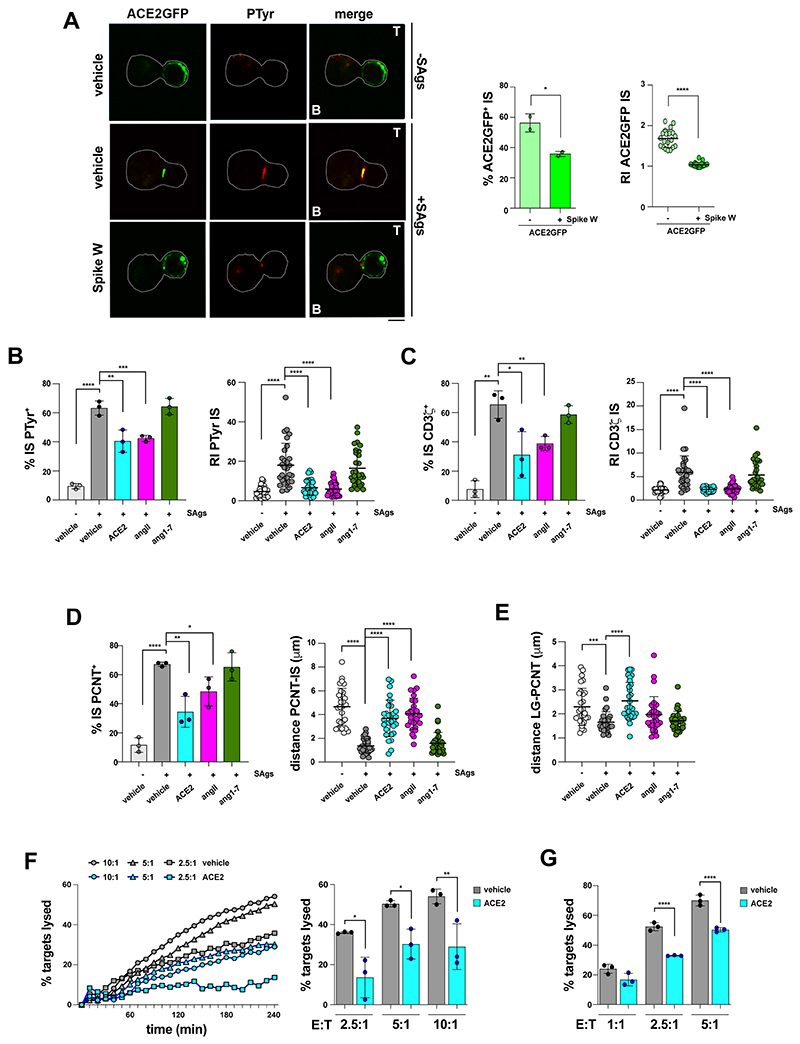
Figure 5. ACE2 is recruited to the CTL IS and suppresses IS formation and cytotoxicity.
A. Immunofluorescence analysis of ACE2 localization in purified CD8+ T cells transfected with an ACE2-GFP-expressing construct or empty GFP vector, pre-treated with either vehicle (PBS) or 0.05 μg/μl Spike Wuhan (Spike W), then mixed with Raji cells (APCs) either unpulsed or pulsed with a combination of SEA, SEB and SEE (SAgs), and incubated for 15 min at 37°C. Representative images are shown. The histograms show (left) the quantification (%) of conjugates harboring ACE2-GFP staining at the IS (≥50 cells/sample, n=2, One-way ANOVA test, *p≤0.05), or (right) the relative ACE2-GFP fluorescence intensity at the IS (recruitment index) (10 cells/sample, n=2, Kruskal-Wallis test, ****p≤0.0001). B,C. Left, Quantification (%) of 15-min conjugates harboring PTyr (B) or CD3ζ (C) staining at the IS. CTLs (day 7) were conjugated with Raji cells (APCs) in the absence or presence of SAgs and either an anti-ACE2 Ab (cell viability after pre-treatment 93.7±0.8%), or angiotensin II (AngII), or the peptide angiotensin 1-7 (Ang 1-7) (≥50 cells/sample, n=3, One-way ANOVA test, ****p≤0.0001; *** p≤0.001; **p≤0.01; *p≤0.05). Right, Relative PTyr (B) or CD3ζ (C) fluorescence intensity at the IS (recruitment index) (10 cells/sample, n=3, Kruskal-Wallis test, ****p≤0.0001). D. Left, Quantification (%) of 15-min conjugates formed as in panel A harboring PCTN staining at the IS (≥50 cells/sample, n=3, One-way ANOVA test, ****p≤0.0001; **p≤0.01; *p≤0.05). Right, Measurement of the distance (μm) of the centrosome (PCNT) from the T cell-APC contact site in CTL-APC conjugates formed as in panel A (10 cells/sample, n=3, Kruskal-Wallis test, ****p≤0.0001). E. Measurement of the distance (μm) of the lytic granules (LG, marked by GzmB) from the centrosome (PCNT) in 15-min CTL-APC conjugates formed as in panel A (10 cells/sample, n=3, Kruskal-Wallis test, ****p≤0.0001; *** p≤0.001). F. Fluorimetric analysis of cytotoxicity of CTLs (day 7) using the calcein release assay. CTLs were pre-treated with either vehicle (PBS) or 2 μg/ml anti-ACE2 Ab (ACE2) and co-cultured with SAg-pulsed, calcein AM-loaded Raji cells at different E:T cell ratios for 4 h. The representative curves show the kinetics of target cell lysis by CTLs at the indicated E:T cell ratios. The histogram shows the target cell lysis at 4 h (n=3, One-way ANOVA test, **p≤0.01; *p≤0.05). G. Flow cytometric analysis of antigen-specific target cell killing by melanoma-specific CTLs derived from 3 patients, using as target the melanoma cell line A375. CTLs were pre-treated as in panel F, co-cultured with A375 cells for 18 h and processed for flow cytometry after staining with propidium iodide and anti-CD8 mAb. Analyses were carried out gating on CD8-/PI+ cells. The histograms show the percentage (%) of target cells lysed (n=3, One-way ANOVA test, ****p≤0.0001). The data are expressed as mean±SD.. Non-significant differences are not shown. Size bar, 5 μm.

