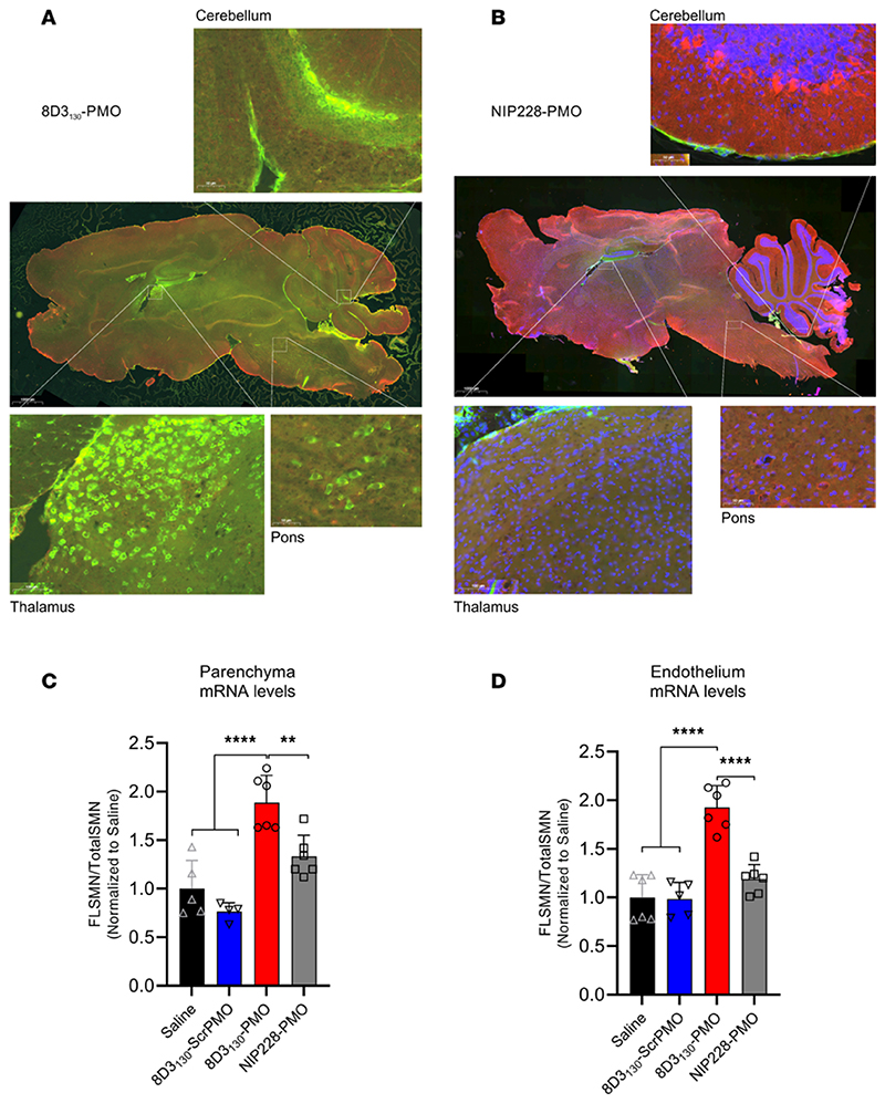
Figure 5. Whole-brain biodistribution of antibody-ASO conjugates.
Representative images of SMN2-transgenic mouse brain treated with single 50 mg/kg administration of (A) 8D3130-PMO or (B) NIP228-PMO. The CNS was isolated from adult mice 24 hours postadministration following perfusion fixation. 8D3130-PMO and NIP228-PMO were identified by human secondary antibody, IgG(H+L). Whole-brain slides were imaged at original magnification 20× on 3DHistech PANNORAMIC 250 slide scanner. Images represent n = 3 mice. The greatest level of 8D3130-PMO uptake into the brain was observed in the thalamus, pons, and cerebellum regions of the brain. (C and D) FLSMN2 expression via qPCR was analyzed in endothelium (BBB) and parenchyma of the brain fractionated by EC extraction. Mice were treated with 50 mg/kg 8D3130-PMO, NIP228-PMO, or 8D3130-scrPMO or 0.9% saline. Statistical significance (representative P values) was evaluated in GraphPad Prism. Data shown as the mean ± SD, n = 6 per group. Results analyzed with 1-way ANOVA corrected for multiple comparisons using Tukey’s test.

