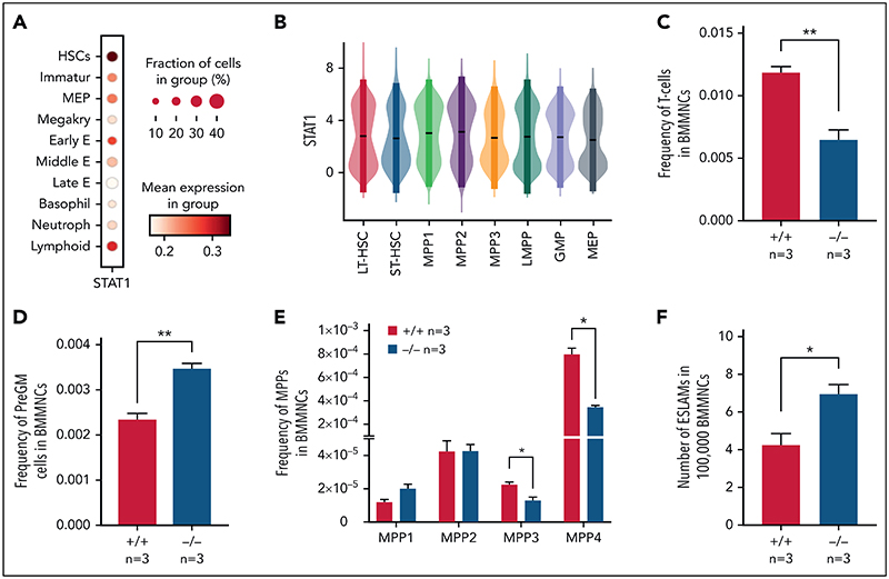
Figure 1. Loss of STAT1 affects the stem and progenitor compartment in steady-state hematopoiesis.
(A) Dot plot showing normalized STAT1 expression in cell types across the Dahlin landscape. The size of each dot indicates the proportion of cells with normalized expression level >0, and the color intensity shows the levels of STAT1 expression. (B) Violin plots showing normalized STAT1 expression in immature cell types in Nestorowa's scRNA-seq dataset. Mean ± standard deviation indicated in overlaid box. (C) The frequency of T cells was reduced in STAT1-deficient bone marrow. (D) The frequency of pre-granulocyte-macrophage progenitors (PreGM) was increased in STAT1-deficient bone marrow. Flow cytometry was performed, and PreGM progenitors were defined as Lin-Sca1-cKit+CD41-CD16/32-CD105-CD150-. (E) The frequencies of MPP3 and MPP4 were reduced in STAT1-deficient bone marrow. Flow cytometry was performed, and multipotent progenitor MPPs were defined as the following: MPP1 (Flk2-CD150+CD48-LSK), MPP2 (Flk2-CD150+CD48+LSK), MPP3 (Flk2-CD150-CD48+LSK), and MPP4 (Flk2+CD150-CD48+LSK). (F) The frequency of ESLAM HSCs was increased in STAT1-deficient mice. Bone marrow ESLAM HSCs were defined as CD45+CD150+CD48-EPCR+ cells. Data are shown as mean ± standard error; asterisks indicate significant differences by Student t test (*P < .05; **P < .01).

