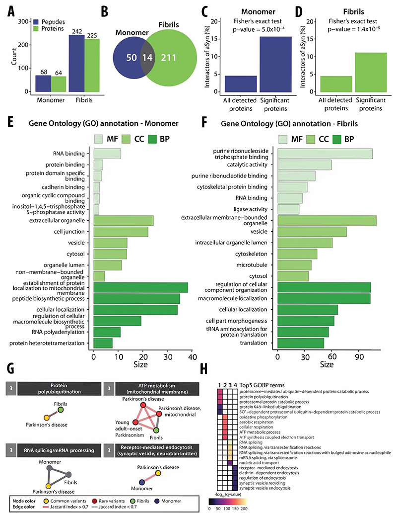
Figure 4. A systematic investigation of structure-specific interactors of the amyloidogenic protein aSyn and gene module analysis of Parkinson’s disease.
(A) Barplot with the numbers of altered LiP peptides (blue) and corresponding structurally altered proteins (green) for aSyn monomer (left) and amyloid fibrils (right). (B) Venn diagram with the numbers of structurally altered proteins for aSyn monomer (blue) and for amyloid fibrils (green). The overlap of structurally altered proteins identified for both aSyn monomer and amyloid fibrils is indicated in gray. (C) The plots show the fraction of known aSyn interactors (based on the STRING database) in structurally altered proteins (right) versus all detected proteins (left) upon spike-in of aSyn monomer into an iPSC-derived cortical neuron extract. The p value assessing enrichment (Fisher’s exact test) is shown. (D) Enrichment plot as in (C) upon spike-in of aSyn fibrils. (E, F) Functional enrichment analyses of structurally altered proteins upon spike-in of aSyn monomer (E) or fibril (F), based on the indicated ontologies (molecular function in light green, cellular component in green, biological process in dark green); the plots show the size (i.e., a score calculated based on q value) of the top6 significant gene ontology terms upon removal of redundant terms (q value <0.01, Benjamini–Hochberg FDR, minimum hypergeometric test, SimRel functional similarity, size = 0.7). (G) Identified modules with enriched GOBP (q value <0.05, Fisher’s exact test, one-sided) that are linked to either common (yellow node) or rare variants (red node) of PD genes for aSyn monomer (blue) or fibrils (green). The thickness of lines represents the Jaccard index (red for Jaccard index >0.7, gray for Jaccard index <0.7). (H) Heatmap showing the top4 GOBP terms within each module as indicated in (B). The gradient color indicates the significance based on the results of a GOBP enrichment test (purple = low significance, yellow = high significance).

