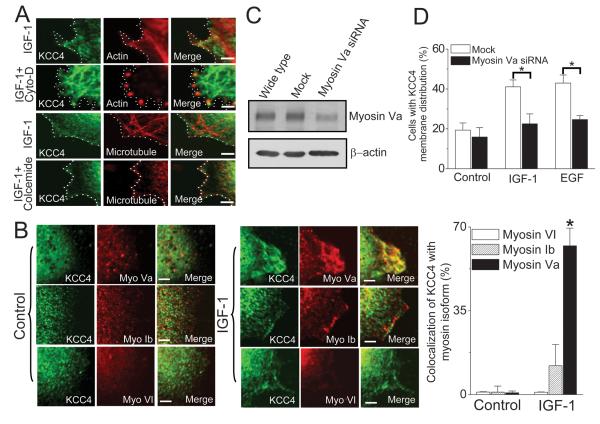
Figure 4. Motor protein-dependent KCC4 trafficking.
(A) KCC4 recruitment is along actin cytoskeleton. Representative confocal images of KCC4 and cytoskeleton in IGF-1-stimulated ovarian cancer OVCAR-3 cells. Cells were pre-incubated with cytochalasin D (cyto-D, 1 μg/ml) or colcemide (10 μg/ml) for 60 min to disrupt actin filaments or microtubule, respectively, prior to 100 ng/ml IGF-1 stimulation. Dashed line: cell periphery. Scale bar, 2 μm. (B) Myosin Va motor protein powers KCC4 membrane trafficking. Left panel: Confocal images of KCC4 and actin-associated motors, such as myosin Va (myo Va), myosin Ib (myo Ib) and myosin VI (myo VI), in the absence (control) or presence of IGF-1 stimulation. Right panel: the colocalization ratio between KCC4 and actin-associated motors at juxta-plasma membrane area with the pixel-by-pixel analyses. Scale bar, 2μm. (C) & (D): The effects of Myosin Va knockdown on growth factor (100 ng/ml)-stimulated KCC4 surface expression. Each column represents mean ± S.E.M. of at least 150 cells. *P<0.01 by unpaired t test.

