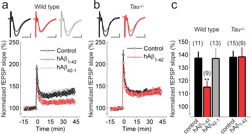
Figure 2. Human Aβ1-42 does not reduce LTP in slices of Tau−/− mice.
(a, b) Hippocampal Schaffer collateral-CA1 LTP in wild-type (a) and Tau−/− mice (b) in control ACSF (black), or after incubation with hAβ1-42 (red) or the control peptide hAβ42-1 (gray). The insets show superimposed example traces before and 40 min after high-frequency stimulation for each condition. Scale bars: 5 ms, 200 μV. (c) Summary of results 40-45 min after high-frequency stimulation. Error bars are s.e.m. ** P < 0.01. The numbers of slices are shown in parentheses.

