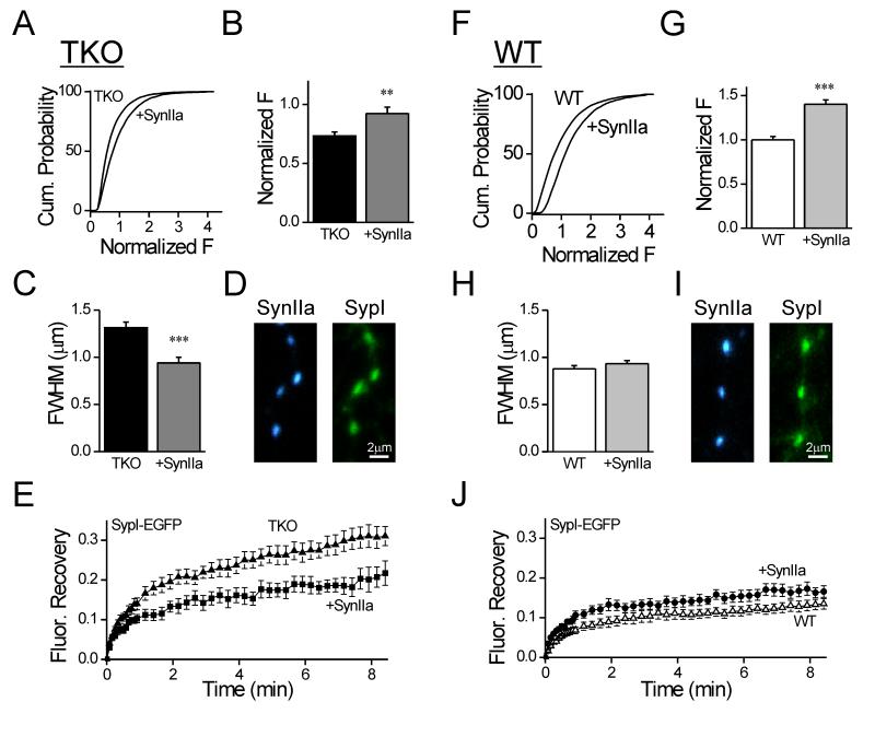
Figure 9. Synapsin IIa rescues the immobilization and clustering of vesicles in synapsin TKO neurons.
(A) Cumulative plot of synaptobrevin 2 immunofluorescence in synaptic puncta of TKO neurons expressing EGPF-synapsin IIa in comparison to uninfected neurons. (B) Average of the means of synaptobrevin 2 signal calculated for independent images. EGFP-synapsin IIa increases the intensity of synaptobrevin 2 staining. The data was normalized by the mean intensity observed in synapses of wildtype neurons (i.e. panel G). (C) Average FWHM width of the synaptobrevin 2 signal in synaptic puncta in TKO neurons expressing EGFP-synapsin IIa in comparison to uninfected neurons. (D) Representative images of synapses in TKO neurons co-expressing TagBFP-Synapsin IIa (blue, left) and SypI-EGFP (green, right). (E) Effect of co-expression of TagBFP-Synapsin IIa (filled squares) on the FRAP time course of SypI-EGFP in TKO neurons (filled triangles). TagBFP-Synapsin IIa reduced the mobility of the vesicles. (F-J) Same as (A-E) except performed in WT neurons. Synapsin IIa increased the intensity of synaptobrevin 2 puncta in WT neurons, but did not affect their width, or the time course of FRAP. ** p<0.01, *** p<0.001.

