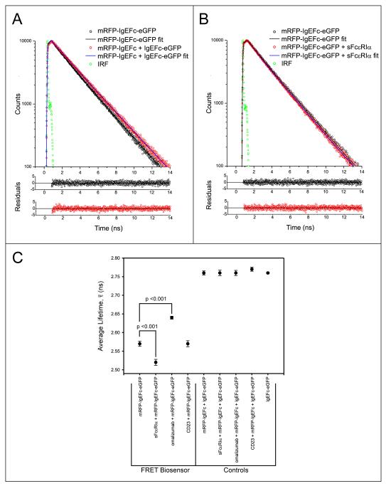
FIG. 2.
Fluorescence lifetime measurements for biosensor and control molecules in the presence and absence of IgE ligands. A: Fluorescence decays (excitation 468nm, emission 510nm) and bi-exponential fits (line plots) and their photon-counting weighted residuals for the mRFP-IgEFc-eGFP biosensor (open black circles), or control molecules (open red circles). Green circles are the instrument response function (IRF). Non-overlap of the fits indicates a shorter fluorescence lifetime for the mRFP-IgEFc-eGFP biosensor, and that it is therefore undergoing FRET. B: Fluorescence decays (excitation 468nm, emission 510nm) and bi-exponential fits (line plots) and their photon-counting weighted residuals, for the mRFP-IgEFc-eGFP biosensor alone (open black circles), or saturated with sFcεRIα (open red circles). Green circles are the instrument response function (IRF). Non-overlap of the fits indicates a shorter fluorescence lifetime for the mRFP-IgEFc-eGFP biosensor in the presence of sFcεRIα. C: Mean values of the results of independent experiments (n = 5) for the true average lifetimes calculated using eqn 2. Significant values were determined using a one-way ANOVA with Tukey-Kramer test. Only the biosensor mRFP-IgEFc-eGFP shows FRET, with changes in true average lifetimes on binding to both sFcεRIα and omalizumab Fab.

