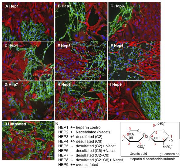
Figure 3. HS sulfation is critical for boundary formation.
Confrontation assays of OECs and astrocytes were carried out in the presence of 10 μg/ml modified heparins (A-J). The disaccharide structures of the heparins are indicated. After 2 days of treatment, cells were fixed and stained for GFAP (red) and p75NTR (green). Assays were scored using a graded scoring system to assess the ability of each modified heparin to promote boundary formation, i.e., mingling (−), partial boundary (+/−) or complete boundary (++) (K). Control heparin effect was scored as ++. Modified heparins with the highest levels of sulfation (Hep1, Hep2, Hep3, Hep4 and Hep9) induced boundaries between OECs and astrocytes, whereas those with lower levels of sulfation (Hep5, Hep6, Hep7 and Hep8) did not. The images are representative of at least 3 independent experiments. Scale bar 50 μm.

