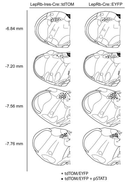
FIG. 1.
Neuroanatomical distribution and validation of transgenically labeled LepRb-expressing neurons of the NTS. LepRb-expressing neurons in LepRb-Ires-Cre::tdTOM and LepRb-Cre::EYFP mice were found across the rostral-caudal extent of the NTS with the preponderance being localized to the NTS at the level of the area postrema. The validity of reporter expression in each line was confirmed via leptin-induced pSTAT3-IR and colocalization with tdTOM- or EYFP-expressing neurons. Each dot ( ) represents five reporter-labeled neurons negative for pSTAT3 and each star (★) five reporter-labeled neurons positive for pSTAT3. AP, Area postrema; cc, central canal; 4v, fourth ventricle.

