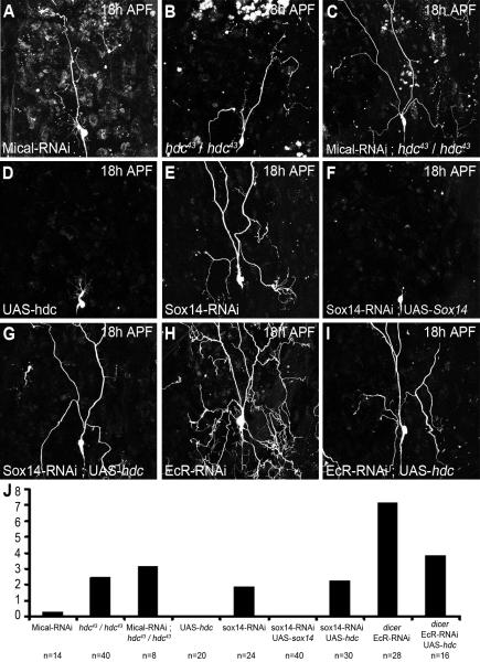
Figure 7. Epistasis experiments between hdc, Sox14 and EcR.
ddaC neurons labeled with UAS-CD8-GFP driven by ppk-GAL4 at 18h APF, also used to drive Mical-RNAi, UAS-hdc, Sox14-RNAi, UAS-Sox14, Mical-RNAi and EcR-RNAiCA104 alone or in combination.
A, the expression of Mical-RNAi can lead to severing defect in 20% of ddaC neurons. B, severing defect observed in 80% of flies homozygous for hdc43. C, the expression of Mical-RNAi in ddaC neurons of homozygous hdc43 flies has an additive effect with 100% of the ddaC neurons presenting severing defect. D, hdc gain-of-function does not affect pruning of ddaC neurons. E, F, the strong severing defect observed with the expression of Sox14-RNAi (E) is completely rescued by the over expression of UAS-Sox14 (F). G, The expression of UAS-hdc is not sufficient to rescue the severing defect generated by Sox14-RNAi. H, I, The almost complete suppression of pruning in ddaC neuron expressing EcR-RNAiCA104 (H) is partially rescued by the over expression of hdc (I). J, Quantification of I° and II° dendrites remaining attached to the soma. There is no significant difference between Sox14-RNAi and the combination of Sox14-RNAi and UAS-hdc. Although not total, the rescue of EcR-RNAiCA104 phenotype by UAS-hdc is significant (P=<0.0001).

