Figure 7.
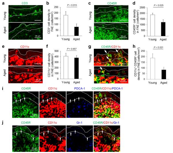
The density of CD11c+ B cells in the FAE of aged mice is significantly reduced. (a, c, e & g) IHC comparison of the distribution of T cells (panel a; CD3+ cells; green), B cells (panel c; CD45R+ cells, green), CD11c+ cells (panel e; red) and CD11c+ B cells (panel g, i and j) in the FAE of young and aged mice. Arrows in panel g indicate CD11c+ B cells. (b, d, f & g) Morphometric analysis of the density of T cells (panel b; CD3+ cells), B cells (panel d; CD45R+ cells), CD11c+ cells (panel f) and CD11c+ B cells (panel h) in the FAE of young and aged mice. (I & j) IHC analysis confirmed that the CD11c+ CD45R+ cells were not plasmacytoid DC as they lacked expression of the typical plasmacytoid DC markers PDCA-1 (blue, panel i) and Gr-1 (blue, panel j). Data are derived from 3-5 Peyer’s patches from 4 mice from each group.

