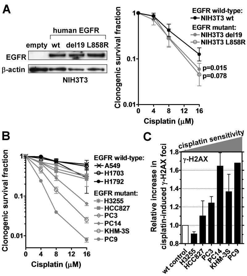
Figure 1.
Cisplatin sensitivity of EGFR-mutant cell lines. A, Left panel, Western blot demonstrating expression of human EGFR in NIH3T3 MEFs transfected with wild-type (wt) or mutant E746_A750 (del19) or L858R constructs. Right panel, Clonogenic survival of MEF clones after 1 hour treatment with cisplatin. B, Clonogenic survival of lung cancer cell lines analogous to panel A. C, Relative increase in the fraction of cells with ≥20 γ–H2AX foci 24 hours after cisplatin treatment (8 μM). EGFR-mutant cell lines are ranked according to their relative cisplatin sensitivity with wild-type (wt) A549 as control. All data show mean ± standard error based on 2-3 biological repeats.

