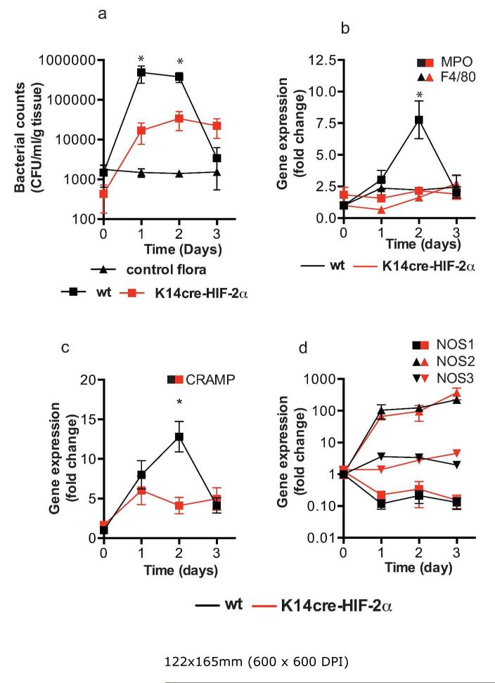
Figure 4. K14cre-HIF-2α mice show reduced wound environmental bacterial flora and reduced acute inflammatory responses.
(a) Bacterial flora (CFU/mg tissue) were calculated from the wounds of K14cre-HIF-2α (n=4) and wild type control (n=4) during the initial 3 days post wounding, and compared to normal background bacterial flora (control). (b and c) QPCR analysis (fold change) of primers for MPO, F4/80 and CRAMP showed a significantly reduced signal in the wounds from K14cre-HIF-2α (n=4) on day 2 when compared to wild type control (n=4) (d) QPCR analysis (fold change) of NOS1-3 showed little deviation between K14cre-HIF-2α (n=4) and wild type controls (n=4).

