Fig. 1.
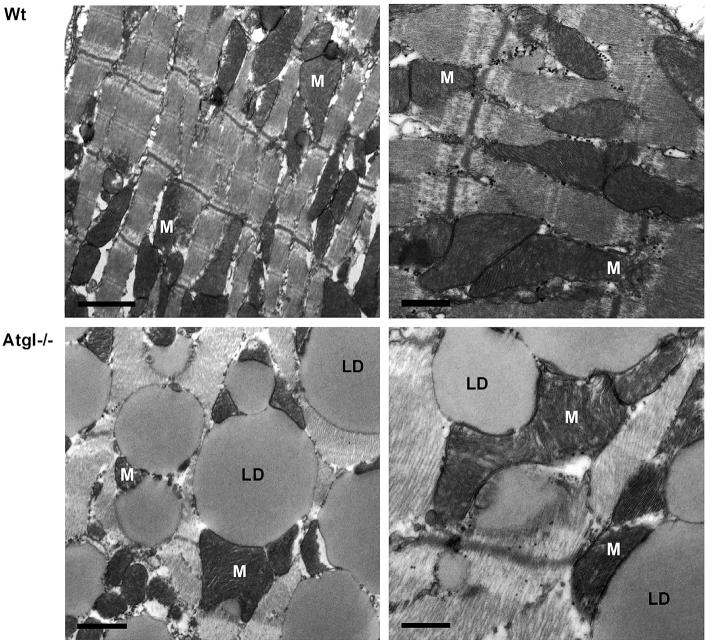
Transmission electron micrographs of heart sections from Wt and Atgl−/− mice. Heart tissue was dissected using a Zeiss OPI1 surgical microscope (Carl Zeiss, Oberkochen, Germany). Small tissue fragments were fixed in 2.5% glutaraldehyde and 2% paraformaldehyde in 0.1 M phosphate buffer (pH 7.4) for 2 h, postfixed in 2% osmium tetroxide for 2 h at room temperature, dehydrated in graded series of ethanol, and embedded in a TAAB epoxy resin. Sections (70 nm thick) were contrasted with uranyl acetate and lead citrate. Images were taken using an FEI Tecnai G2 20 transmission electron microscope (FEI Eindhoven, Eindhoven, Netherlands) with a Gatan ultrascan 1000 CCD camera. Acceleration voltage used was 120 kV. Wt cardiac muscle sections (upper panels) show a intermyofibrillar network containing mitochondria (M). Atgl−/− cardiac muscle (lower panels) show a massive lipid droplet (LD) accumulation within the intermyofibrillar network. Overall morphology of mitochondria and the structure of mitochondrial cristae appeared normal. Mitochondrial size compared to that of Wt mice is increased [14]. Left panels: scale bars 1 μm; right panels: scale bars 0.5 μm.

