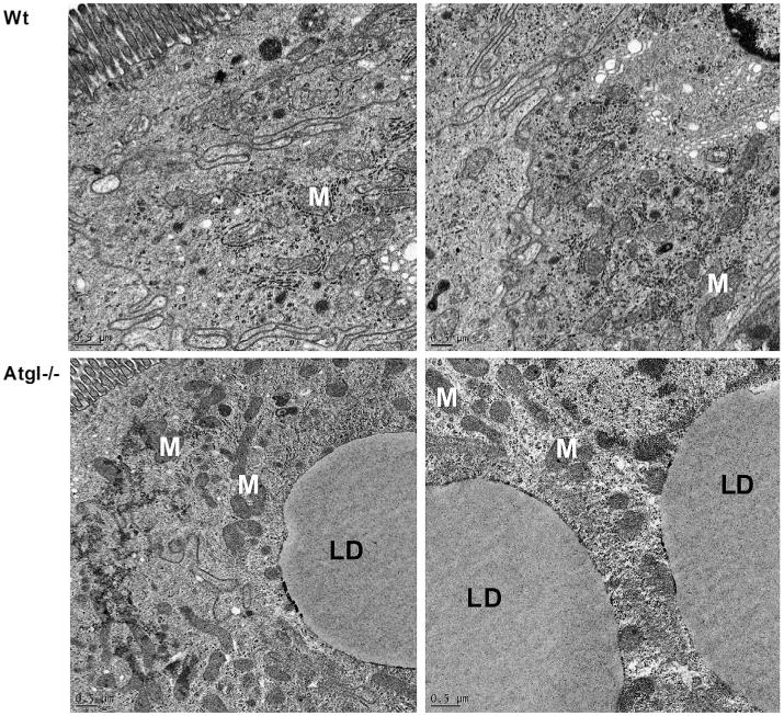
Fig. 4.
Transmission electron micrographs of small intestinal sections from Wt and intestine-specific Atgl−/− mice. Section from Wt (upper panels) and Atgl−/− (lower panels) small intestinal cells show intact mitochondria (M) albeit lipid droplet (LD) accumulation in Atgl−/− small intestine. Scale bars: 0.5 μm.

