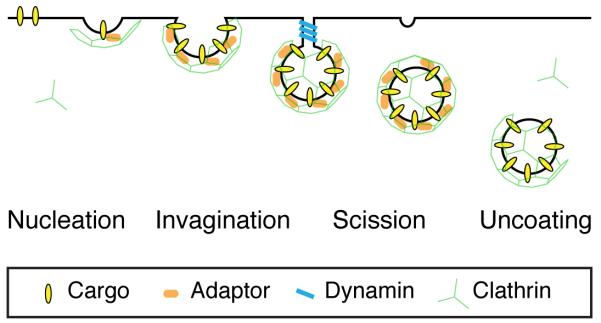
Figure 1. Schematic diagram of clathrin-mediated endocytosis.

The four main stages of clathrin-mediated endocytosis (CME) are shown. 1. Nucleation, where cargo is gathered into the forming pit. 2. Invagination, where the pit curves inwards. 3. Scission, where the CCV is severed from the plasma membrane. 4. Uncoating, where the clathrin coat is removed and the nascent vesicle is freed. The basic layer of molecules that drive this process is shown as indicated. The second layer of proteins that add further complexity to the pathway are not shown.
