
Figure 3.
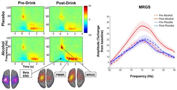
Grand-averaged time-frequency spectrograms of motor responses for each condition and a map of grand-averaged source activity across all conditions shown on MNI template brains. For MRGS responses a power by frequency plot of average non-fitted data is presented with shaded within-subject error. There is a clear alcohol-induced increase in amplitude.