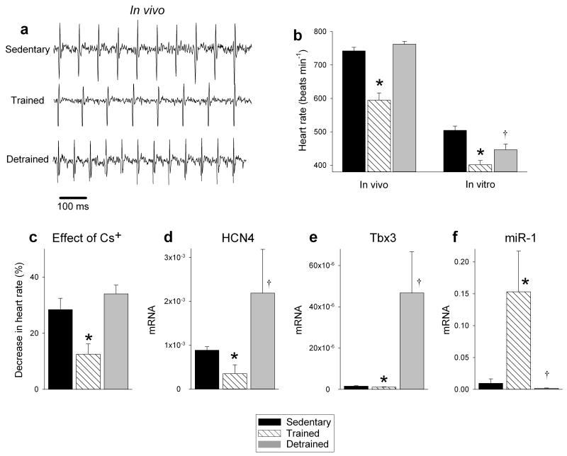
Figure 5. Sinus bradycardia and sinus node remodelling in the mouse are reversed by detraining.
a, Representative ECG traces obtained from sedentary, trained and detrained mice (unrestrained and conscious). b, Mean (+SEM) heart rate measured in vivo (n=7/8/5) and in vitro (from isolated sinus node preparations; n=7/8/5) in sedentary, trained and detrained mice. c, Restoration of the contribution of If to pacemaking in the detrained mouse. Mean (+SEM) percentage decrease in heart rate of isolated sinus node preparations from sedentary, trained and detrained mice on blocking If using 2 mM CsCl shown (n=6/7/4). d-f, Restoration of normal levels of HCN4 and Tbx3 mRNA and miR-1 in the sinus node in the detrained mouse. Mean (+SEM) expression of HCN4 and Tbx3 mRNA (normalised to 18S) and miR-1 (normalised to RNU1A1) in the sinus node of sedentary, trained and detrained mice shown (n=4). Student’s t test used to test differences. Normal distribution of data was tested using the Shapiro-Wilk W test and equal variance was tested using the F test. When the null hypothesis of normality and/or equal variance was rejected, the nonparametric Mann Whitney U test was used. *P<0.05, trained versus sedentary mice; †P<0.05, detrained versus trained mice.

