Fig. 1.
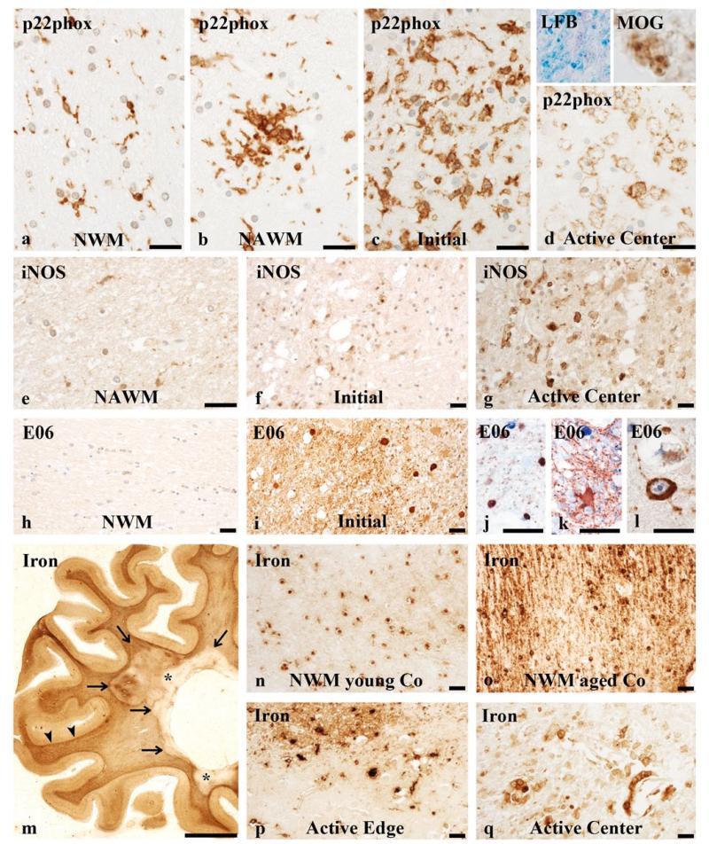
Oxidative burst, oxidative injury and iron accumulation in MS lesions. Expression of p22phox in the normal white matter (NWM) of a control brain (a), the normal-appearing white matter (NAWM; b), the zone of initial (pre-phagocytic) demyelination (c) and in the plaque centre of an active lesion (d) from an acute MS patient, defined by the presence of LFB and MOG reactive myelin degradation products in macrophages (inserts). Individual microglia in the control brain expressed p22phox. There was profound microglial activation in the NAWM with the formation of microglial nodules, a massive p22phox expression in initial areas of active demyelination and a low expression on macrophages containing myelin debris in the plaque centre. iNOS expression in acute MS in the NAWM (e) and in initial areas of demyelination (f) was weak and restricted to a few cells with microglial morphology. In the active plaque centre, iNOS was mainly expressed on macrophages (g). Immunoreactivity of E06 staining for oxidised phospholipids was low or absent in the NWM of controls (h) and very high in areas of initial demyelination in acute MS (i). Double staining for E06 (blue) and cell markers (brown) documented its presence in oligodendrocytes (stained for TPPPp25; j) and cytoplasmic granules in astrocytes (stained for GFAP; k). Intense E06 reactivity was also found in the cytoplasm of cortical neurons, showing beading and fragmentation of their cell processes (l). Iron staining of the MS brain revealed profound iron accumulation in the subcortical white matter (m; arrowheads) and at the edge of active lesions (m; arrows) from a SPMS patient. Established lesions showed a reduced iron staining (m; asterisks). Young control brains revealed a relatively low iron content mainly found in oligodendrocytes (n; age 30). The aged human control (o; age 84) and MS (m; age 57) brains displayed high amounts of non-haeme iron in oligodendrocytes and myelin. High iron content was present at the edge of an active lesion from a RRMS patient in microglia (p). Within the centre of an active lesion, the iron load was reduced and iron reactivity was mainly found in (perivascular) macrophages (q). Scale bar 50 μm except for m = 1 cm

